 Explore
 Validate
 Learn
Learn Immunohistochemistry
 Other assay
Antibody data
- Antibody Data
- Antigen structure
- References [0]
- Comments [0]
- Validations
- Other assay [2]
Submit
Validation data
Reference
Comment
Report error
- Product number
- MA1-35420 - Provider product page

- Provider
- Invitrogen Antibodies
- Product name
- HLA-DR Monoclonal Antibody (LN-3)
- Antibody type
- Monoclonal
- Antigen
- Other
- Description
- The antibody reacts with human leukocyte antigen-DR (HLA-DR). The mAb has lymph node germinal center and mantle zone B cell reactivity. It reacts with interdigitating histiocytes in T cell zones and with sinus histiocytes and endothelial cells. It has also tumor specificity and reactivity with normal non-lymphoid tissue.
- Reactivity
- Human
- Host
- Mouse
- Isotype
- IgG
- Antibody clone number
- LN-3
- Vial size
- 1 mL
- Concentration
- 250 µg/mL
- Storage
- 4° C
No comments: Submit comment
Supportive validation
- Submitted by
- Invitrogen Antibodies (provider)
- Main image

- Experimental details
- NULL

- Submitted by
- Invitrogen Antibodies (provider)
- Main image
- Experimental details
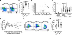
- Fig 2 Memory distributions, activation, and proportion of cytotoxic peripheral CD8 + T cells for healthy donors and following HIV infection. ( A ) Representative flow cytometric plots of CCR7 versus CD45RO from a pre- (day -159) and acute (day 28) infection time point for one donor. ( B ) Proportion of circulating total memory CD8 + T cells for all healthy donors (HD; n = 41), acute HIV time points (23-100 d; n = 27), and chronic HIV time points (>365 d; n = 23). ( C ) Peak viral load plotted against total memory CD8 + T cells at the earliest available time point post-infection (23-41 d) for each RV217 donor. Spearman's rank correlation test was used to determine significance. ( D ) Memory subsets as determined by CCR7 and CD45RO staining for all healthy donors (circles), acute HIV time points (squares), and chronic HIV time points (triangles). ( E ) Proportion of memory CD8 + T cells that express HLA-DR over time from infection for four RV217 subjects. Pre-infection time points were set as day 0 for analysis. ( F ) Representative flow cytometric plots of perforin and HLA-DR expression by memory CD8 + T cells from a pre- (day -173), acute (day 31), and chronic (day 420) infection time point for one donor. SS Day 420 sample was acquired and analyzed at a later date than earlier samples resulting in a different gating scheme. For consistency, gates were set using naive (CCR7 + CD45RO - ) CD8 + T cells, which generally do not express perforin or HLA-DR. ( G ) Proportion of
