 Explore
 Validate
 Learn
Learn Western blot
 Immunocytochemistry
Antibody data
- Antibody Data
- Antigen structure
- References [0]
- Comments [0]
- Validations
- Western blot [1]
- Immunohistochemistry [3]
- Flow cytometry [3]
Submit
Validation data
Reference
Comment
Report error
- Product number
- NBP2-47670-0.1mg - Provider product page

- Provider
- Novus Biologicals
- Product name
- Mouse Monoclonal HLA-DR Antibody
- Antibody type
- Monoclonal
- Description
- Protein A or G purified. This MAb reacts with a 28kDa chain of HLA-DRB1 antigen, a member of MHC class II molecules. It does not cross react with HLA-DP and HLA-DQ. The L243 antibody recognizes a different epitope than the LN3 monoclonal antibody, and these antibodies do not cross-block binding to each other's respective epitopes. HLA-DR is a heterodimeric cell surface glycoprotein comprised of a 36kDa alpha (heavy) chain and a 28kDa beta (light) chain. It is expressed on B-cells, activated T-cells, monocytes/macrophages, dendritic cells and other non-professional APCs. In conjunction with the CD3/TCR complex and CD4 molecules, HLA-DR is critical for efficient peptide presentation to CD4+ T cells. It is an excellent histiocytic marker in paraffin sections producing intense staining. True histiocytic neoplasms are similarly positive. HLA-DR antigens also occur on a variety of epithelial cells and their corresponding neoplastic counterparts. Loss of HLA-DR expression is related to tumor microenvironment and predicts adverse outcome in diffuse large B-cell lymphoma.
- Reactivity
- Human, Mouse, Simian
- Host
- Mouse
- Isotype
- IgG
- Vial size
- 0.1 mg
- Concentration
- 1.0 mg/ml
- Storage
- Store at 4C short term. Aliquot and store at -20C long term. Avoid freeze-thaw cycles.
No comments: Submit comment
Supportive validation
- Submitted by
- Novus Biologicals (provider)
- Main image
- Experimental details
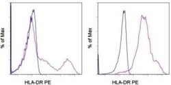
- Western Blot: HLA-DR Antibody (LN-3) - Azide and BSA Free [NBP2-47670] - Analysis using the PE conjugate of NBP2-34676. Staining of normal human peripheral blood cells with Mouse IgG2b K Isotype Control PE (blue histogram) or Anti-Human HLA-DR PE (purple histogram). Cells in the lymphocyte (left) or monocyte (right) gate were
Supportive validation
- Submitted by
- Novus Biologicals (provider)
- Main image
- Experimental details
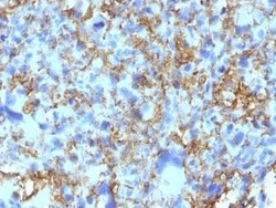
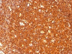
- Immunohistochemistry-Paraffin: HLA-DR Antibody (LN-3) - Azide and BSA Free [NBP2-47670] - Human Histiocytoma stained with HLA-DRB Monoclonal Antibody (LN-3).
- Submitted by
- Novus Biologicals (provider)
- Main image
- Experimental details
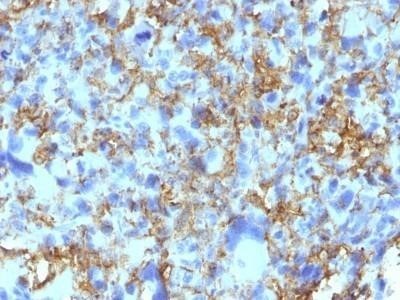
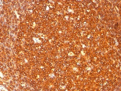
- Immunohistochemistry-Paraffin: HLA-DR Antibody (LN-3) - Azide and BSA Free [NBP2-47670] - Analysis using Azide and BSA Free version of NBP2-47670. Formalin-paraffin human tonsil stained with HLA-DR MAb (LN-3).
- Submitted by
- Novus Biologicals (provider)
- Main image
- Experimental details
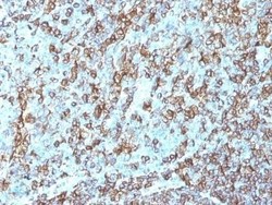
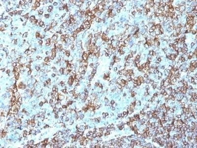
- Immunohistochemistry-Paraffin: HLA-DR Antibody (LN-3) - Azide and BSA Free [NBP2-47670] - Human Tonsil stained with HLA-DRB Monoclonal Antibody (LN-3).
Supportive validation
- Submitted by
- Novus Biologicals (provider)
- Main image

- Experimental details
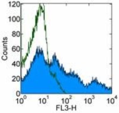
- Flow Cytometry: HLA-DR Antibody (LN-3) - Azide and BSA Free [NBP2-47670] - Analysis using the PerCP/Cy5.5 conjugate of NBP2-34676. Staining of normal human peripheral blood cells with Mouse IgG2b kappa Isotype Control PerCP-Cy5.5 (open histogram) or Anti-Human HLA-DR PerCP-Cy5.5 (filled histogram). Cells in the lymphocyte gate were used for analysis.
- Submitted by
- Novus Biologicals (provider)
- Main image

- Experimental details
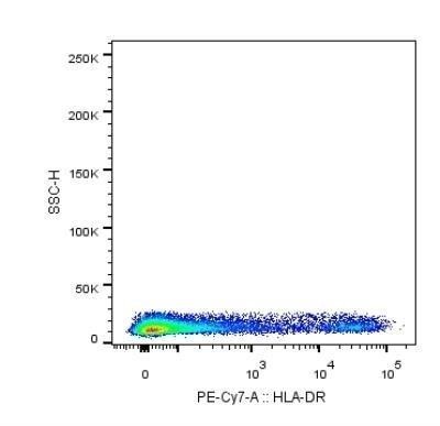
- Flow Cytometry: HLA-DR Antibody (LN-3) - Azide and BSA Free [NBP2-47670] - Analysis using the PE/Cy7 conjugate of NBP2-34676. Staining of HLA-DR in human buffy coat using anti-HLA-DR antibody. The primary antibody was used at a dilution of 1:100 and incubated for 25 min at 4C in 2% human serum, 0.5mM EDTA in DPBS. Image from verified customer review.
- Submitted by
- Novus Biologicals (provider)
- Main image
- Experimental details
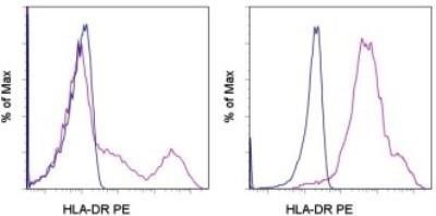
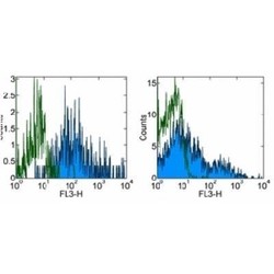
- Flow Cytometry: HLA-DR Antibody (LN-3) - Azide and BSA Free [NBP2-47670] - Analysis using the PE/Cy7 conjugate of NBP2-34676. Staining of normal human peripheral blood cells with staining buffer (autofluorescence) (open histogram) or Anti-Human HLA-DR PE-Cyanine7 (filled histogram). Cells in the lymphocyte (right) or monocyte (left) gate were used for analysis.
