 Explore
 Validate
 Learn
Learn37-5900
antibody from Invitrogen Antibodies
Targeting: NECTIN1
CD111, CLPED1, ED4, HIgR, HVEC, OFC7, PRR, PRR1, PVRL1, PVRR1, SK-12
 Western blot
 ELISA
ELISAAntibody data
- Antibody Data
- Antigen structure
- References [8]
- Comments [0]
- Validations
- Western blot [3]
- Other assay [10]
Submit
Validation data
Reference
Comment
Report error
- Product number
- 37-5900 - Provider product page

- Provider
- Invitrogen Antibodies
- Product name
- Nectin 1 Monoclonal Antibody (CK8)
- Antibody type
- Monoclonal
- Antigen
- Recombinant full-length protein
- Reactivity
- Human, Mouse, Rat
- Host
- Mouse
- Isotype
- IgG
- Antibody clone number
- CK8
- Vial size
- 100 µg
- Concentration
- 0.5 mg/mL
- Storage
- -20°C
Submitted references Newly Characterized Murine Undifferentiated Sarcoma Models Sensitive to Virotherapy with Oncolytic HSV-1 M002.
Nonmuscle myosin heavy chain IIb mediates herpes simplex virus 1 entry.
HSV-1 infection of human corneal epithelial cells: receptor-mediated entry and trends of re-infection.
A novel function of heparan sulfate in the regulation of cell-cell fusion.
Role of nectin-1, HVEM, and PILR-alpha in HSV-2 entry into human retinal pigment epithelial cells.
HVEM and nectin-1 are the major mediators of herpes simplex virus 1 (HSV-1) entry into human conjunctival epithelium.
The keratin-binding protein Albatross regulates polarization of epithelial cells.
The host adherens junction molecule nectin-1 is downregulated in Chlamydia trachomatis-infected genital epithelial cells.
Ring EK, Li R, Moore BP, Nan L, Kelly VM, Han X, Beierle EA, Markert JM, Leavenworth JW, Gillespie GY, Friedman GK
Molecular therapy oncolytics 2017 Dec 15;7:27-36
Molecular therapy oncolytics 2017 Dec 15;7:27-36
Nonmuscle myosin heavy chain IIb mediates herpes simplex virus 1 entry.
Arii J, Hirohata Y, Kato A, Kawaguchi Y
Journal of virology 2015 Feb;89(3):1879-88
Journal of virology 2015 Feb;89(3):1879-88
HSV-1 infection of human corneal epithelial cells: receptor-mediated entry and trends of re-infection.
Shah A, Farooq AV, Tiwari V, Kim MJ, Shukla D
Molecular vision 2010 Nov 20;16:2476-86
Molecular vision 2010 Nov 20;16:2476-86
A novel function of heparan sulfate in the regulation of cell-cell fusion.
O'Donnell CD, Shukla D
The Journal of biological chemistry 2009 Oct 23;284(43):29654-65
The Journal of biological chemistry 2009 Oct 23;284(43):29654-65
Role of nectin-1, HVEM, and PILR-alpha in HSV-2 entry into human retinal pigment epithelial cells.
Shukla SY, Singh YK, Shukla D
Investigative ophthalmology & visual science 2009 Jun;50(6):2878-87
Investigative ophthalmology & visual science 2009 Jun;50(6):2878-87
HVEM and nectin-1 are the major mediators of herpes simplex virus 1 (HSV-1) entry into human conjunctival epithelium.
Akhtar J, Tiwari V, Oh MJ, Kovacs M, Jani A, Kovacs SK, Valyi-Nagy T, Shukla D
Investigative ophthalmology & visual science 2008 Sep;49(9):4026-35
Investigative ophthalmology & visual science 2008 Sep;49(9):4026-35
The keratin-binding protein Albatross regulates polarization of epithelial cells.
Sugimoto M, Inoko A, Shiromizu T, Nakayama M, Zou P, Yonemura S, Hayashi Y, Izawa I, Sasoh M, Uji Y, Kaibuchi K, Kiyono T, Inagaki M
The Journal of cell biology 2008 Oct 6;183(1):19-28
The Journal of cell biology 2008 Oct 6;183(1):19-28
The host adherens junction molecule nectin-1 is downregulated in Chlamydia trachomatis-infected genital epithelial cells.
Sun J, Kintner J, Schoborg RV
Microbiology (Reading, England) 2008 May;154(Pt 5):1290-1299
Microbiology (Reading, England) 2008 May;154(Pt 5):1290-1299
No comments: Submit comment
Supportive validation
- Submitted by
- Invitrogen Antibodies (provider)
- Main image
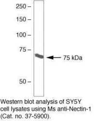
- Experimental details
- Western blot analysis of SY5Y cell lysates using Ms anti-Nectin-1 (Product # 37-5900)

- Submitted by
- Invitrogen Antibodies (provider)
- Main image
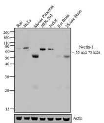
- Experimental details
- Western blot analysis of Nectin-1 was performed by loading 20 µg of Raji (lane1), HeLa (lane2), Mouse Pancreas (lane3), HEK-293 (lane4), Jurkat (lane5), Rat Brain (lane6) and Mouse Brain (lane7) lysate using Novex®NuPAGE® 12 % Bis-Tris gel (Product # NP0342BOX), XCell SureLock Electrophoresis System (Product # EI0002), Novex® Sharp Pre-Stained Protein Standard (LC5800), and iBlot® 2 Dry Blotting System (IB21001). Proteins were transferred to a nitrocellulose membrane and blocked with 5% skim milk for 1 hour at room temperature. Nectin-1 was detected at ~ 55 kDa in Rodent tissue lysates and at ~ 75 kDa in Human cell lysates using Nectin-1 Mouse Monoclonal Antibody (Product # 37-5900) at 0.1-1 µg/mL in 5% skim milk at 4°C overnight on a rocking platform. Goat Anti-Mouse - HRP Secondary Antibody (Product # 62-6520) at 1:4000 dilution was used and chemiluminescent detection was performed using Pierce™ ECL Western Blotting Substrate (Product # 32106).

- Submitted by
- Invitrogen Antibodies (provider)
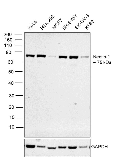
- Main image

- Experimental details
- Western blot was performed using Anti-Nectin 1 Monoclonal Antibody (CK8) (Product # 37-5900) and a 75 kDa band corresponding to Nectin-1 was observed across all the cells liens tested. Whole-cell extracts (30 µg lysate) of HeLa (Lane 1), HEK293 (Lane 2), MCF7 (Lane 3), SH-SY5Y (Lane 4), SK-OV-3 (Lane 5), K562 (Lane 6) were electrophoresed using NuPAGE™ 4-12% Bis-Tris Protein Gel (Product # NP0322BOX). Resolved proteins were then transferred onto a nitrocellulose membrane (Product # IB23001) by iBlot® 2 Dry Blotting System (Product # IB21001). The blot was probed with the primary antibody (0.5 µg/mL) and detected by chemiluminescence with Goat anti-Mouse IgG (H+L) Superclonal™ Recombinant Secondary Antibody, HRP (Product # A28177,1:4000 dilution) using the iBright™ FL1500 Imaging System (Product # A44115). Chemiluminescent detection was performed using SuperSignal™ West Dura Extended Duration Substrate (Product # 34076).
Supportive validation
- Submitted by
- Invitrogen Antibodies (provider)
- Main image

- Experimental details
- NULL
- Submitted by
- Invitrogen Antibodies (provider)
- Main image
- Experimental details
- NULL
- Submitted by
- Invitrogen Antibodies (provider)
- Main image
- Experimental details
- NULL
- Submitted by
- Invitrogen Antibodies (provider)
- Main image
- Experimental details
- NULL
- Submitted by
- Invitrogen Antibodies (provider)
- Main image

- Experimental details
- NULL

- Submitted by
- Invitrogen Antibodies (provider)
- Main image
- Experimental details
- NULL
- Submitted by
- Invitrogen Antibodies (provider)
- Main image
- Experimental details
- NULL
- Submitted by
- Invitrogen Antibodies (provider)
- Main image
- Experimental details
- NULL

- Submitted by
- Invitrogen Antibodies (provider)
- Main image
- Experimental details
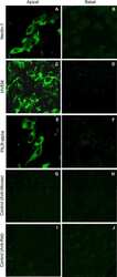
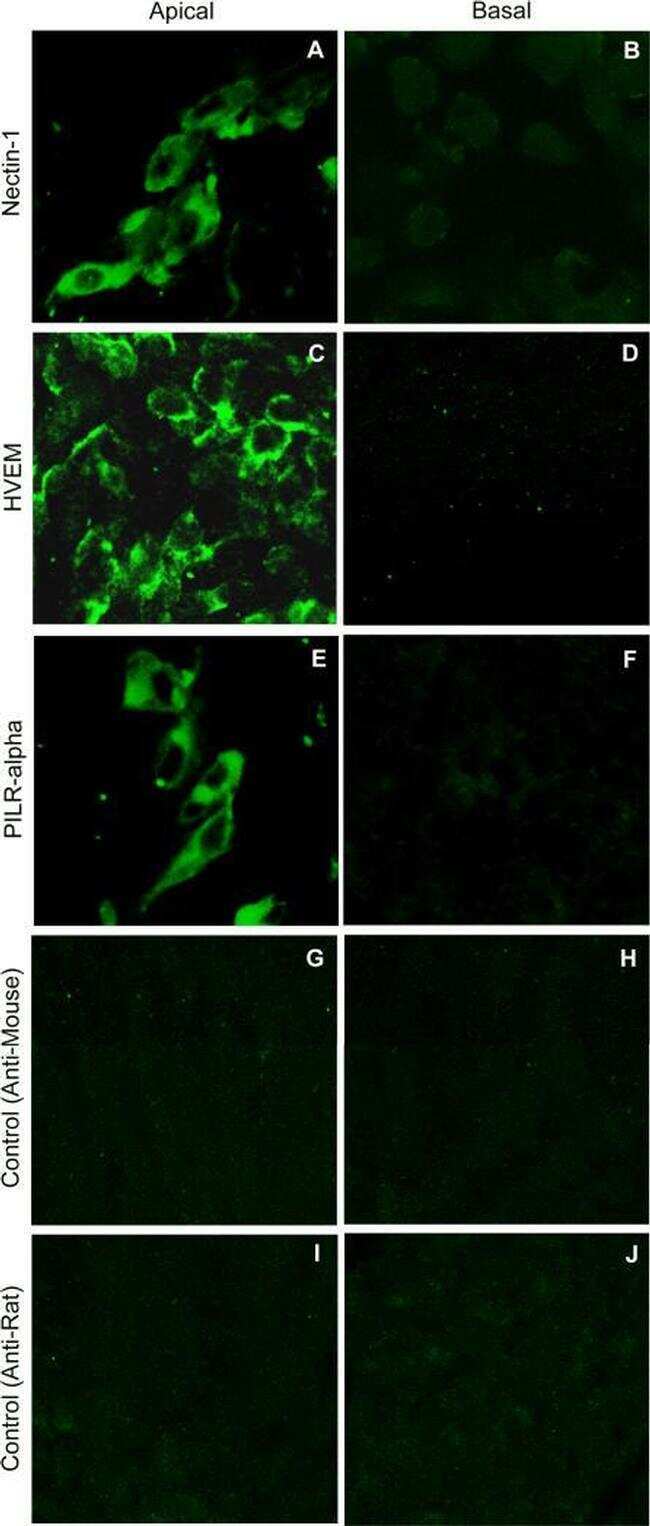
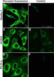
- Figure 5 Immunofluorescence imaging of receptors on HCE cell membrane. Images shown were taken using the FITC filter of confocal microscope (Leica SP20). Cells were blocked for 90 min, washed, and then either mock treated with buffer alone ( B , D , F ) or treated with primary antibodies for Nectin-1 ( A ), HVEM ( C ), and PILR-alpha ( E ). Images were taken after the incubation of HCE cells with FITC-conjugated secondary antibodies. Staining of cells with green demonstrate receptor expression.

- Submitted by
- Invitrogen Antibodies (provider)
- Main image
- Experimental details
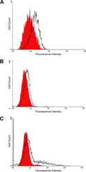
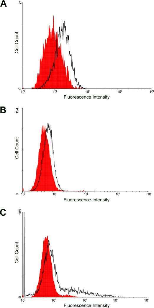
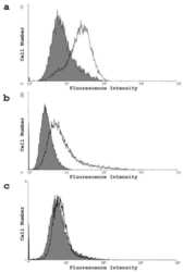
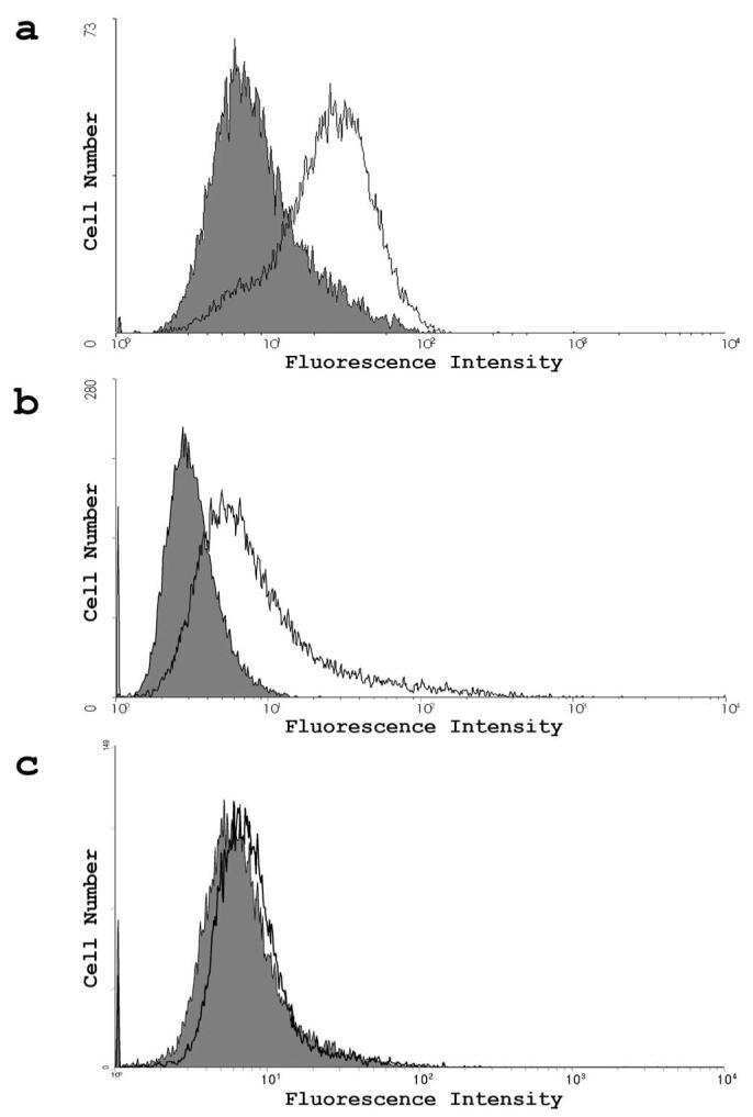
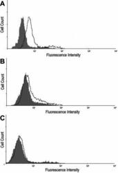
- Figure 6 Flow cytometry analysis of cell-surface receptor expression. Expression was detected by Fluorescence-activated cell sorter (FACS) analysis. Cells were treated with primary antibodies to Nectin-1 ( A ), HVEM ( B ), or PILR-alpha ( C ). HCE cells stained only with FITC-conjugated secondary antibody were used as background controls and are shown as the dark gray in the figure.
