 Explore
 Validate
 Learn
Learn Western blot
 Immunoprecipitation
Antibody data
- Antibody Data
- Antigen structure
- References [0]
- Comments [0]
- Validations
- Western blot [5]
- Other assay [1]
Submit
Validation data
Reference
Comment
Report error
- Product number
- 711820 - Provider product page

- Provider
- Invitrogen Antibodies
- Product name
- Parkin Recombinant Polyclonal Antibody (21HCLC)
- Antibody type
- Polyclonal
- Antigen
- Other
- Reactivity
- Human, Mouse, Rat
- Host
- Rabbit
- Isotype
- IgG
- Antibody clone number
- 21HCLC
- Vial size
- 100 µg
- Concentration
- 0.5 mg/mL
- Storage
- Store at 4°C short term. For long term storage, store at -20°C, avoiding freeze/thaw cycles.
No comments: Submit comment
Supportive validation
- Submitted by
- Invitrogen Antibodies (provider)
- Main image

- Experimental details
- Western blot analysis was performed on Whole cell extracts (30 µg lysate) of SH-SY5Y (Lane 1), Neuro-2a (Lane 2) and tissue extracts of Mouse Brain (Lane 3) and Rat Brain (Lane 4). The blots were probed with Anti-Parkin Recombinant Rabbit Polyclonal Antibody (Product # 711820, 2.5 µg/mL) and detected by chemiluminescence using Goat anti-Rabbit IgG (H+L) Superclonal™ Secondary Antibody, HRP conjugate (Product # A27036, 0.25 µg/mL, 1:4000 dilution). A 48 kDa band corresponding to Parkin was observed across the cell lines and tissues tested. Known quantity of protein samples were electrophoresed using Novex®NuPAGE®4-12% Bis-Tris gel (Product # NP0322BOX), XCell SureLock™ Electrophoresis System (Product # EI0002) and Novex® Sharp Pre-Stained Protein Standard (Product # LC5800). Resolved proteins were then transferred onto a nitrocellulose membrane with iBlot® Dry Blotting System (Product # IB21001). The membrane was probed with the relevant primary and secondary Antibody following blocking with 5% skimmed milk. Chemiluminescent detection was performed using Pierce™ ECL Western blotting Substrate (Product # 32106).

- Submitted by
- Invitrogen Antibodies (provider)
- Main image
- Experimental details
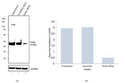
- Knockdown of Parkin was achieved by transfecting SH-SY5Y cells with Parkin specific siRNA (Silencer® select product # S10043+s10045). Western blot analysis was performed using membrane extracts from the Parkin knock down cells (lane 3), non-specific scrambled siRNA transfected cells (lane 2) and untransfected cells (lane 1). The blots were probed with Anti-Parkin Recombinant Rabbit Polyclonal Antibody (Product # 711820, 1-3 µg/mL) and Goat anti-Rabbit IgG (H+L) Superclonal™ Secondary Antibody, HRP conjugate (Product # A27036, 0.4 µg/mL, 1:2500 dilution). Loss of signal upon siRNA mediated knock down confirms that antibody is specific to Parkin.

- Submitted by
- Invitrogen Antibodies (provider)
- Main image

- Experimental details
- Western blot analysis was performed on Whole cell extracts (30 µg lysate) of SH-SY5Y (Lane 1), Neuro-2a (Lane 2) and tissue extracts of Mouse Brain (Lane 3) and Rat Brain (Lane 4). The blots were probed with Anti-Parkin Recombinant Rabbit Polyclonal Antibody (Product # 711820, 2.5 µg/mL) and detected by chemiluminescence using Goat anti-Rabbit IgG (H+L) Superclonal™ Secondary Antibody, HRP conjugate (Product # A27036, 0.25 µg/mL, 1:4000 dilution). A 48 kDa band corresponding to Parkin was observed across the cell lines and tissues tested. Known quantity of protein samples were electrophoresed using Novex®NuPAGE®4-12% Bis-Tris gel (Product # NP0322BOX), XCell SureLock™ Electrophoresis System (Product # EI0002) and Novex® Sharp Pre-Stained Protein Standard (Product # LC5800). Resolved proteins were then transferred onto a nitrocellulose membrane with iBlot® Dry Blotting System (Product # IB21001). The membrane was probed with the relevant primary and secondary Antibody following blocking with 5% skimmed milk. Chemiluminescent detection was performed using Pierce™ ECL Western blotting Substrate (Product # 32106).

- Submitted by
- Invitrogen Antibodies (provider)
- Main image
- Experimental details
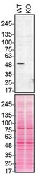
- Western blot of Parkin was performed by loading 50 µg of WT (lane 1) and PRKN CRISPR KO (lane 2) SH-SY5Y cell lysates in RIPA buffer onto a 4-15% gradient polyacrylamide gel. Proteins on the blots were visualized with Ponceau staining (below immunoblot). Proteins were transferred to nitrocellulose membrane and blocked in 5% milk for 1 hr. PRKN was detected at approximately 52 kDa using a PRKN recombinant polyclonal antibody (Product # 711820) at a dilution of 1:200 in 5% BSA in TBS with 0.1% Tween 20 (TBST) overnight at 4°C. The peroxidase-conjugated secondary antibody (Product # 65-6120) was diluted to 0.2 µg/mL in TBST with 5% milk for 1 hr. Chemiluminescent detection was performed using Pierce ECL Western Blotting Substrate (Product # 32106). Data courtesy of YCharOS Inc., an open science company with the mission of characterizing commercially available antibodies using knockout validation.

- Submitted by
- Invitrogen Antibodies (provider)
- Main image
- Experimental details
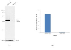
- Western blot analysis (Fig a) of Parkin was performed on cell extracts (100 µg of lysate) of HEK-293 wild type (Lane 1) and Parkin knockout (Lane 2). The blot was probed with Anti-Parkin Recombinant Rabbit Polyclonal Antibody (Product # 711820, 1:500 dilution) and detected by chemiluminescence using Peroxidase AffiniPure Goat anti-Rabbit IgG (H+L) Secondary Antibody, HRP conjugate (Product # 111-035-144, 1:4000 dilution). Densitometric analysis of this Western blot is shown in the histogram (Fig b). Loss of signal upon CRISPR mediated knockout (KO) confirms that antibody is specific to Parkin.

Supportive validation
- Submitted by
- Invitrogen Antibodies (provider)
- Main image

- Experimental details
- Immunoprecipitation of PRKN was performed on SH-SY5Y cell lysates. Antibody-bead conjugates were prepared by adding 1 µg of PRKN recombinant polyclonal antibody (Product # 711820) with 30 µL of protein A-Sepharose beads and rocked overnight at 4°C. 2 mg of lysate was incubated with an antibody-bead conjugate for 2 hours at 4°C. Following centrifugation and multiple washes, 10% starting material (SM), 10% unbound fraction (UB) and immunoprecipitated fraction (IP) were processed for immunoblot using another PRKN monoclonal antibody (Product # 39-0900). Ponceau stained transfer of blot is shown. Data courtesy of YCharOS Inc., an open science company with the mission of characterizing commercially available antibodies using knockout validation.
