Explore
 Validate
 Learn
Learn Immunohistochemistry
 Other assay
Antibody data
- Antibody Data
- Antigen structure
- References [0]
- Comments [0]
- Validations
- Other assay [2]
Submit
Validation data
Reference
Comment
Report error
- Product number
- 16-9243-85 - Provider product page

- Provider
- Invitrogen Antibodies
- Product name
- TGF beta-1,2,3 Monoclonal Antibody (1D11.16.8), Functional Grade, eBioscience™
- Antibody type
- Monoclonal
- Antigen
- Other
- Description
- Description: The monoclonal antibody 1D11.16.8 recognizes human, mouse, rat, hamster, canine and non-human primate Transforming Growth Factor (TGF) beta 1, 2 and 3. TGF beta is a pleiotropic cytokine that exists as five isoforms (TGF beta 1-5) that share 70-80% homology with each other. However, these isoforms are not homologous to TGF alpha. TGF beta 1 is highly conserved between human, murine, simian, bovine, porcine, and chicken proteins. TGF beta 1 is the most abundant isoform and is ubiquitously expressed by a variety of cell types, including platelets, macrophages, lymphocytes, endothelial cells, chondrocytes, and leukemic cells. However, the other isoforms exhibit a more restricted expression pattern. TGF beta 1 is synthesized as a long precursor polypeptide, which is cleaved to yield the mature protein and the Latency Associated Peptide (LAP). LAP and mature TGF beta 1 remain non-covalently associated during secretion, forming homodimers known as the Small Latent Complex (SLC). Secretion can be induced by steroids, retinoids, EGF, NGF, vitamin D3, and IL-1. The bioactivity of mature TGF beta 1 is dependent on its release from LAP by conformational changes and proteolytic processing. Its activities include inhibition of cell growth in epithelial cells, endothelial cells, fibroblasts, neurons, lymphoid cells, and other hematopoietic cell types. TGF beta 1 also inhibits the proliferation of T and NK cells, downregulates the activities of activated macrophages, and blocks the anti-tumor activity of IL-2 - bearing lymphokine-activated killer (LAK) cells. Recently, TGF beta 1 has been found to play a critical role in the development of regulatory T cells and act as a costimulatory factor for expression of Foxp3. Dendritic cells exposed to tumors have been reported to secrete TGF beta 1 and stimulate the differntiation of CD4+CD25+ Treg cell from peripheral CD4+CD25- progeny. TGF beta 1-induced regulatory T cells have been termed iTregs. Applications Reported: This 1D11.16.8 antibody has been reported for use in neutralization assays both in vivo and in vitro and immunohistochemical staining of formalin-fixed paraffin embedded tissue sections. Storage and handling: Use in a sterile environment. Filtration: 0.2 µm post-manufacturing filtered. Purity: Greater than 90%, as determined by SDS-PAGE. Endotoxin Level: Less than 0.001 ng/µg antibody, as determined by LAL assay. Aggregation: Less than 10%, as determined by HPLC.
- Reactivity
- Human, Mouse, Rat, Canine, Hamster
- Host
- Mouse
- Isotype
- IgG
- Antibody clone number
- 1D11.16.8
- Vial size
- 500 μg
- Concentration
- 1 mg/mL
- Storage
- 4°C
No comments: Submit comment
Supportive validation
- Submitted by
- Invitrogen Antibodies (provider)
- Main image
- Experimental details
- NULL
- Submitted by
- Invitrogen Antibodies (provider)
- Main image
- Experimental details
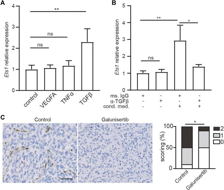
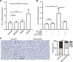
- FIGURE 2 Ets1 upregulation in glioblastoma endothelial cells depend on TGFbeta signaling. (A) RNA levels of Ets1 in bEND.3 cells treated with VEGFA, TNFalpha or TGFbeta (mean + SD of four independent experiments; ** p < 0.01; ANOVA with Tukey's multiple comparisons test). (B) RNA levels of Ets1 in bEND.3 cells treated with conditioned medium from CT-2A glioma cells with TGFbeta neutralizing antibody or control antibody (mean + SD of four independent experiments; ** p < 0.01; One-way ANOVA with Tukey's multiple comparisons test). (C) Immunohistochemical staining and quantification of Ets1 in CT-2A glioma tumor-bearing mice treated with/without galunisertib (bar: 50 mum) ( n = 10/group, Mann-Whitney test, * p < 0.05).