Explore
 Validate
 Learn
Learn ELISA
ELISAAntibody data
- Antibody Data
- Antigen structure
- References [0]
- Comments [0]
- Validations
- ELISA [2]
- Other assay [1]
Submit
Validation data
Reference
Comment
Report error
- Product number
- 13-9923-85 - Provider product page

- Provider
- Invitrogen Antibodies
- Product name
- TGF beta-1,2,3 Monoclonal Antibody (eBio16TFB), Biotin, eBioscience™
- Antibody type
- Monoclonal
- Antigen
- Other
- Description
- Description: The eBio16TFB antibody reacts with Transforming Growth Factor-beta (TGF-beta) a pleiotropic cytokine, exists in five isoforms, known as TGF-ß1-5. Homologies between isoforms range from 70-80% but no homology exists to TGF-a. TGF-ß1 is ubiquitous and the most abundant form found in lymphoid organs, while other isoforms are expressed in a more restricted distribution. The biologically active state of all isoforms are disulfide-linked homodimers. The heat- and acid- stable monomeric subunits have a length of 112 amino acids. The heat- and acid- stable monomeric subunits have a length of 112 amino acids. The isoforms of TGF-beta arise by proteolytic cleavage of longer precursors; the isoforms are derived from the carboxyterminal ends of these precursors. Isoforms isolated from different species are evolutionarily closely conserved and have sequence identities on the order of 98%. Mature human, porcine, simian, chicken and bovine TGF-beta1 are identical and differ from mouse TGF-beta1 in a single amino acid. TGF-beta1 is produced in very high levels by platelets. Other cellular sources of TGF-beta1 include macrophages, lymphocytes, endothelial cells, chondrocytes, and leukemic cells. TGF-beta1 secretion can be induced by steroids, retinoids, EGF, NGF, vitamin D3, and IL-1. Activities of TGF-beta1 include inhibition of cell growth for inhibitor for normal and transformed epithelial cells, endothelial cells, fibroblasts, neurons, and lymphoid cells and other hematopoietic cell types. TGF-beta1 inhibits the proliferation of T cells and NK cells and downregulates the activities of activated macrophages. TGF-beta1 blocks the anti-tumor activity of IL-2 - bearing lymphokine-activated killer (LAK) cells. Recently, TGF-beta1 has been found to have a critical role in the development of regulatory T cells. Dendritic cells exposed to tumors have been reported to secrete TGF-beta1 and stimulate expansion of naturally-occurring T reg cells. Moreover, TGF-beta1 has been shown to act as a costimulatory factor for expression of Foxp3, leading to the differentiation of CD4+CD25+ Treg cells from peripheral CD4+CD25- progeny. TGF-beta-induced regulatory T cells have been termed Ti-Treg. Applications Reported: This eBio16TFB antibody has been reported for use in ELISA. Applications Tested: The eBio16TFB antibody has been tested as the detection antibody in a sandwich ELISA for analysis of human and mouse TGF-beta in combination with the capture eBioTB2F (Product # 14-9943-81) antibody for detection. A suitable range of concentrations of this antibody for ELISA capture is 2-6 µg/mL. A standard curve consisting of doubling dilutions of the recombinant standard over the range of 8000 pg/mL - 60 pg/mL should be included in each ELISA plate. This antibody recognizes the mature/active form of TGF beta 1 without association with Latency Associated Peptide (LAP). Most samples will require acid-treatment and neutralization to remove LAP from TGF beta 1 prior to evaluation in this assay. Samples should be tested in the assay immediately after acid treatment and neutralization. It is also possible that some serum and plasma samples may contain low levels of immunoreactive TGF beta 1 that has disassociated from LAP. Naturally occurring, free TGF beta 1 may be measurable by evaluating samples without acid treatment. Filtration: 0.2 µm post-manufacturing filtered.
- Reactivity
- Human
- Host
- Mouse
- Conjugate
- Biotin
- Isotype
- IgG
- Antibody clone number
- eBio16TFB
- Vial size
- 500 μg
- Concentration
- 0.5 mg/mL
- Storage
- 4°C, do not freeze
No comments: Submit comment
Supportive validation
- Submitted by
- Invitrogen Antibodies (provider)
- Main image

- Experimental details
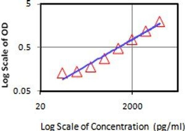
- Standard curve of human/mouse TGF beta ELISA.
- Conjugate
- Biotin

- Submitted by
- Invitrogen Antibodies (provider)
- Main image

- Experimental details
- Standard curve of human/mouse TGF beta ELISA.
Supportive validation
- Submitted by
- Invitrogen Antibodies (provider)
- Main image
- Experimental details
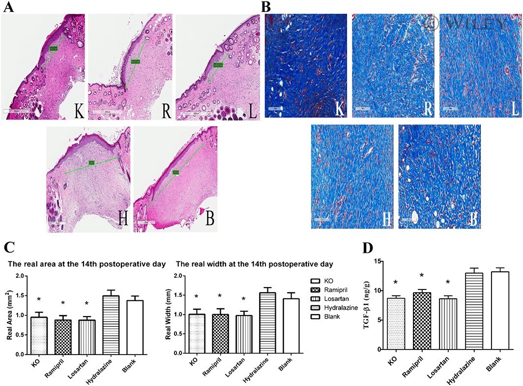
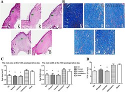
- 2 ACEI reduces scar formation and TGF-beta1 expression on postoperative day 14. (A, B) Typical histological sections of mouse scar tissue obtained on the 14th postoperative day (A: HE staining, B: Masson staining). The green bars (A) are used to mark the scales of scar, the white bars = 500 mum (A) /100 mum (B). In each composite picture, K represents KO group, R represents ramipril group, L represents losartan group, H represents hydralazine group and B represents blank control group. (C) The actual (real) scar area and width were determined in each group of mice ( n = 12 wounds in six mice for each group). The results are given as mean +- SEM. * P < 0.05 as compared to the blank group respectively, by least significant difference (LSD) and Student-Newman-Keuls (SNK) tests (D) The content of TGF-beta1 in 1 g of scar tissue ( n = 12 wounds in six mice for each group). All results are given as mean +- SEM. * P < 0.05 as compared to the blank group, respectively, by LSD and SNK tests.
- Conjugate
- Biotin