Explore
 Validate
 Learn
LearnPA5-21923
antibody from Invitrogen Antibodies
Targeting: H2AZ2
H2AFV, H2AV, MGC10170, MGC10831, MGC1947
 Western blot Immunocytochemistry
 Immunoprecipitation Immunohistochemistry Chromatin Immunoprecipitation Other assay
Antibody data
- Antibody Data
- Antigen structure
- References [0]
- Comments [0]
- Validations
- Western blot [1]
- Immunocytochemistry [3]
- Immunohistochemistry [1]
- Chromatin Immunoprecipitation [2]
- Other assay [1]
Submit
Validation data
Reference
Comment
Report error
- Product number
- PA5-21923 - Provider product page

- Provider
- Invitrogen Antibodies
- Product name
- Histone H2A.Z Polyclonal Antibody
- Antibody type
- Polyclonal
- Antigen
- Synthetic peptide
- Description
- Recommended positive controls: Molt-4, Raji. Predicted reactivity: Mouse (100%), Rat (100%), Xenopus laevis (100%), Dog (100%), Pig (100%), Chicken (100%), Sheep (100%), Rhesus Monkey (100%), Bovine (100%). Store product as a concentrated solution. Centrifuge briefly prior to opening the vial.
- Reactivity
- Human, Mouse
- Host
- Rabbit
- Isotype
- IgG
- Vial size
- 100 µL
- Concentration
- 1 mg/mL
- Storage
- Store at 4°C short term. For long term storage, store at -20°C, avoiding freeze/thaw cycles.
No comments: Submit comment
Supportive validation
- Submitted by
- Invitrogen Antibodies (provider)
- Main image
- Experimental details
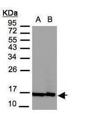
- Western Blot using Histone H2A. Z Polyclonal Antibody (Product # PA5-21923). Sample (30 µg of whole cell lysate). A: MOLT4SDS PAGE. Z. B: Raji. 12% SDS PAGE. Histone H2A. Z Polyclonal Antibody (Product # PA5-21923) diluted at 1:3,000.
Supportive validation
- Submitted by
- Invitrogen Antibodies (provider)
- Main image

- Experimental details
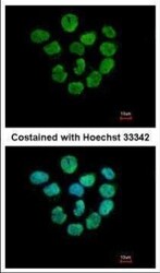
- Immunofluorescent analysis of Histone H2A.Z+H2A.F.Z in paraformaldehyde-fixed A431 cells using a Histone H2A.Z+H2A.F.Z polyclonal antibody (Product # PA5-21923) at a 1:500 dilution.
- Submitted by
- Invitrogen Antibodies (provider)
- Main image
- Experimental details
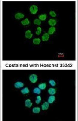
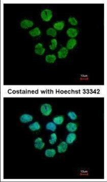
- Immunofluorescence analysis of paraformaldehyde-fixed A431, using Histone H2A. Z antibody (Product # PA5-21923) at 1:500 dilution.
- Submitted by
- Invitrogen Antibodies (provider)
- Main image
- Experimental details
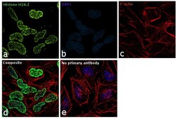
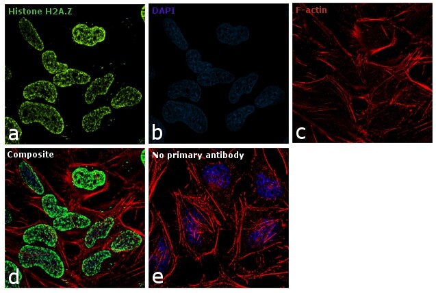
- Immunofluorescence analysis of Histone H2A.Z was performed using 70% confluent log phase HeLa cells. The cells were fixed with 4% paraformaldehyde for 10 minutes, permeabilized with 0.1% Triton™ X-100 for 10 minutes, and blocked with 1% BSA for 1 hour at room temperature. The cells were labeled with Histone H2A.Z Rabbit Polyclonal Antibody (Product # PA5-21923) at 5 µg/mL in 0.1% BSA and incubated overnight at 4 degree and then labeled with Goat anti-Rabbit IgG (H+L) Superclonal™ Secondary Antibody, Alexa Fluor® 488 conjugate (Product # A27034) at a dilution of 1:2000 for 45 minutes at room temperature (Panel a: green). Nuclei (Panel b: blue) were stained with SlowFade® Gold Antifade Mountant with DAPI (Product # S36938). F-actin (Panel c: red) was stained with Rhodamine Phalloidin (Product # R415, 1:300). Panel d represents the merged image showing nuclear localization. Panel e represents control cells with no primary antibody to assess background. The images were captured at 60X magnification.
Supportive validation
- Submitted by
- Invitrogen Antibodies (provider)
- Main image
- Experimental details
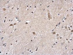
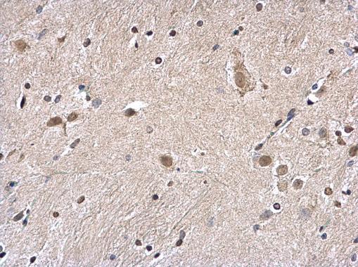
- Histone H2A. Z Polyclonal Antibody detects Histone H2A. Z protein at nucleus on mouse fore brain by immunohistochemical analysis. Sample: Paraffin-embedded mouse fore brain. Histone H2A. Z Polyclonal Antibody (Product # PA5-21923) diluted at 1:1,000. Antigen Retrieval: EDTA based buffer, pH 8.0, 15 min.
Supportive validation
- Submitted by
- Invitrogen Antibodies (provider)
- Main image
- Experimental details
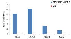
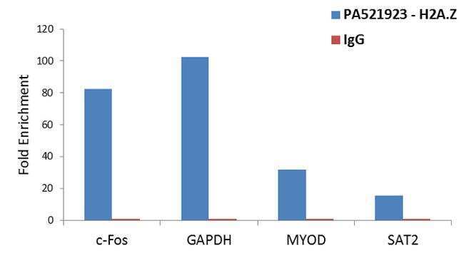
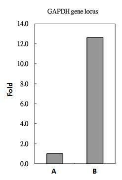
- Enrichment of endogenous Histone H2A.Z protein at specific gene loci using Anti-Histone H2A.Z Antibody: Chromatin Immunoprecipitation (ChIP) was performed using Anti-Histone H2A.Z Rabbit Polyclonal Antibody (Product # PA5-21923, 3 µg) on sheared chromatin from 2 million HeLa cells using the MAGnify ChIP system kit (Product # 49-2024). Normal Rabbit IgG was used as a negative IP control. The purified DNA was analyzed by qPCR with PCR primer pairs for the promoters of c-Fos, GAPDH used as positive and MYOD , SAT2 satellite repeats used as negative target genes/binding sites. Data is presented as fold enrichment of the antibody signal versus the negative control IgG using the comparative CT method.
- Submitted by
- Invitrogen Antibodies (provider)
- Main image
- Experimental details
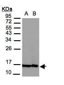
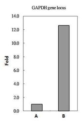
- Histone H2A. Z antibody immunoprecipitates Histone H2A. Z protein-DNA in ChIP experiments. ChIP Sample: HeLa whole cell lysate/extract. A: 5 µg preimmune rabbit IgG. B: 5 µg of Histone H2A. Z antibody (Product # PA5-21923). The precipitated DNA was detected by PCR with primer set targeting to GAPDH gene locus.
Supportive validation
- Submitted by
- Invitrogen Antibodies (provider)
- Main image

- Experimental details
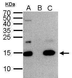
- Histone H2A. Z Polyclonal Antibody immunoprecipitates Histone H2A. Z protein in IP experiments. IP Sample: 1,000 µg HeLa whole cell lysate/extract A. 50 µg HeLa whole cell lysate/extract B. Control with 2 µg of preimmune rabbit IgG C. Immunoprecipitation of Histone H2A. Z protein by 2 µg of Histone H2A. Z Polyclonal Antibody (Product # PA5-21923) 15% SDS-PAGE The immunoprecipitated Histone H2A. Z protein was detected by Histone H2A. Z Polyclonal Antibody (Product # PA5-21923) diluted at 1:1,000.