Explore
 Validate
 Learn
Learn Western blot
Antibody data
- Antibody Data
- Antigen structure
- References [0]
- Comments [0]
- Validations
- Western blot [3]
- Immunohistochemistry [1]
Submit
Validation data
Reference
Comment
Report error
- Product number
- PA5-61600 - Provider product page

- Provider
- Invitrogen Antibodies
- Product name
- RAC1/RAC2/RAC3 Polyclonal Antibody
- Antibody type
- Polyclonal
- Antigen
- Recombinant full-length protein
- Description
- Immunogen sequence: TKLDLRDDKD TIERLRDKKL APITYPQGLA MAREI Highest antigen sequence identity to the following orthologs: Mouse - 100%, Rat - 100%.
- Reactivity
- Human
- Host
- Rabbit
- Isotype
- IgG
- Vial size
- 100 µL
- Concentration
- 0.1 mg/mL
- Storage
- Store at 4°C short term. For long term storage, store at -20°C, avoiding freeze/thaw cycles.
No comments: Submit comment
Supportive validation
- Submitted by
- Invitrogen Antibodies (provider)
- Main image

- Experimental details
- Western blot analysis of RAC1/RAC2/RAC3 in Lane 1: Marker (kDa) 250, 130, 95, 72, 55, 36, 28, 17, 10; Lane 2: Human cell line RT-4. Samples were probed using a RAC1/RAC2/RAC3 Polyclonal Antibody (Product # PA5-61600).

- Submitted by
- Invitrogen Antibodies (provider)
- Main image

- Experimental details
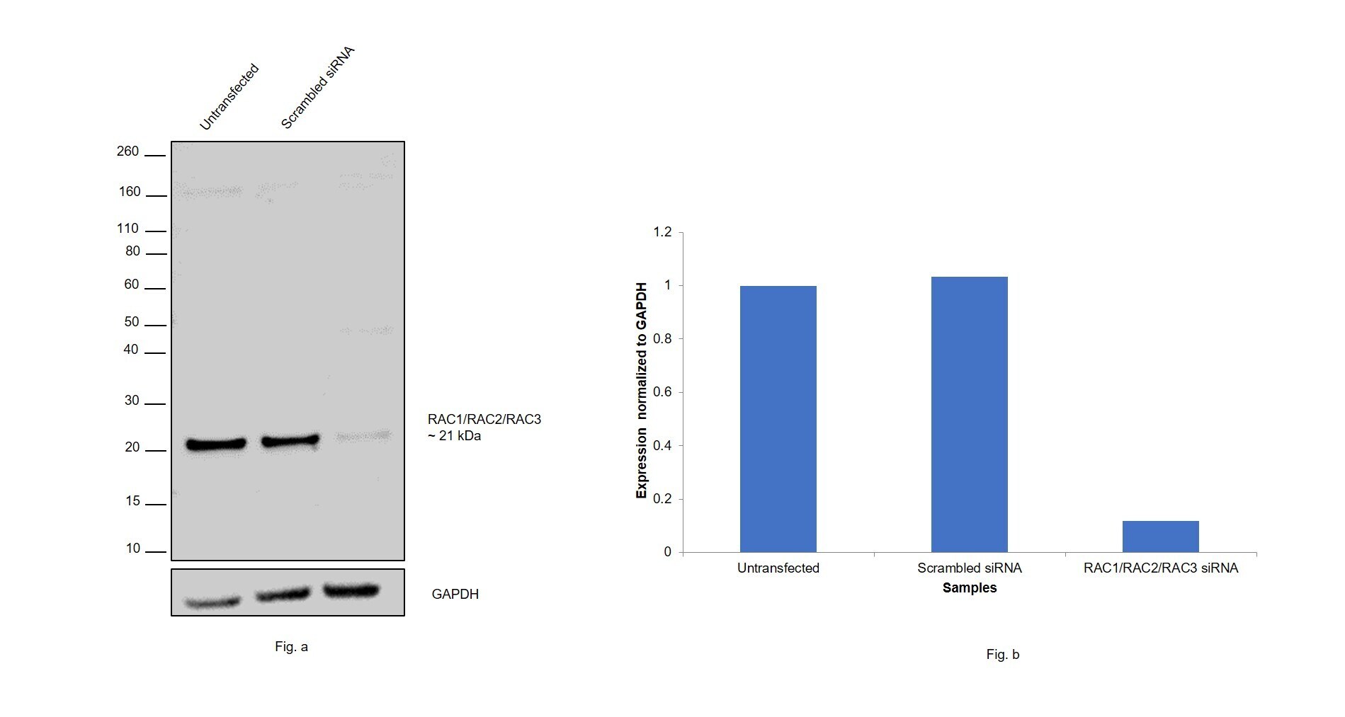
- Knockdown of RAC1/RAC2/RAC3 was achieved by transfecting HEK-293 cells with Rac1/Rac2/Rac3 specific siRNAs (Silencer® select Product # s11713 - RAC1, Product # s11716 & Product # s11714 - RAC2, Product # s11717 & Product # s224513 - RAC3). Western blot analysis (Fig. a) was performed using whole cell extracts from the RAC1/RAC2/RAC3 knockdown cells (lane 3), non-specific scrambled siRNA transfected cells (lane 2) and untransfected cells (lane 1). The blots were probed by RAC1/RAC2/RAC3 Polyclonal Antibody (Product # PA5-61600, 1:500 dilution) and Goat anti-Rabbit IgG (H+L) Superclonal™ Recombinant Secondary Antibody, HRP conjugate (Product # A27036, 0.25 ug/ml, 1:4000 dilution). Densitometric analysis of this western blot is shown in histogram (Fig. b). Decrease in signal upon siRNA mediated knock down confirms that antibody is specific toRac1/Rac2/Rac3.
- Submitted by
- Invitrogen Antibodies (provider)
- Main image

- Experimental details
- Western blot was performed using Anti-RAC1/RAC2/RAC3 Polyclonal Antibody (Product # PA5-61600) and 21 kDa band corresponding to RAC1/RAC2/RAC3 was observed along with some higher glycosylated forms were also seen across samples tested. Whole cell extracts (30 µg lysate) of A-431 (Lane1), Jurkat (Lane2), U-87 MG (Lane 3), HEK-293 (Lane 4), HL-60 (Lane 5), Mouse Brain, (Lane 6), Mouse Thymus (Lane 7) and Mouse Spleen (Lane 8) were electrophoresed using Novex® NuPAGE® 4-12% Bis-Tris gel (Product # NP0342BOX). Resolved proteins were then transferred onto a nitrocellulose membrane (Product # IB23001) by iBlot® 2 Dry Blotting System (Product # IB21001). The blot was probed with the primary antibody (1:500 dilution) and detected by Goat anti-Rabbit IgG (H+L) Superclonal™ Recombinant Secondary Antibody, HRP (Product # A27036, 1:4000 dilution) using the iBright FL 1000 (Product # A32752). Chemiluminescent detection was performed using Novex® ECL Chemiluminescent Substrate Reagent Kit (Product # WP20005).

Supportive validation
- Submitted by
- Invitrogen Antibodies (provider)
- Main image

- Experimental details
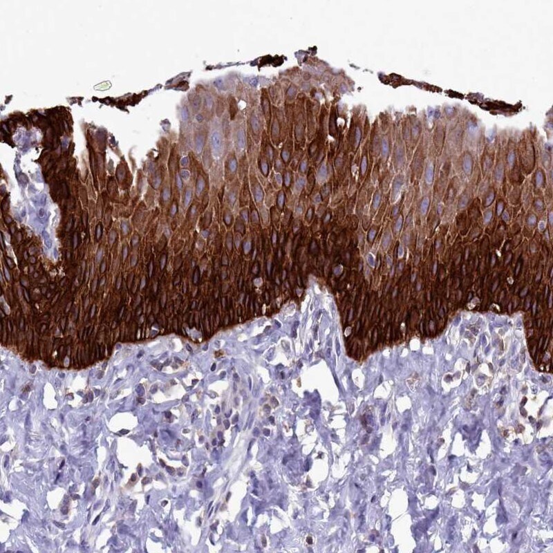
- Immunohistochemical staining of RAC1/RAC2/RAC3 in human esophagus using a RAC1/RAC2/RAC3 Polyclonal Antibody (Product # PA5-61600) shows strong cytoplasmic positivity in squamous epithelial cells.