Explore
 Validate
 Learn
Learn Western blot
Antibody data
- Antibody Data
- Antigen structure
- References [0]
- Comments [0]
- Validations
- Western blot [1]
- Flow cytometry [1]
- Other assay [1]
Submit
Validation data
Reference
Comment
Report error
- Product number
- PA5-17519 - Provider product page

- Provider
- Invitrogen Antibodies
- Product name
- RAC1/RAC2/RAC3 Polyclonal Antibody
- Antibody type
- Polyclonal
- Antigen
- Synthetic peptide
- Description
- It is not recommended to aliquot this antibody. This antibody is not cross-reactive with other small GTPases.
- Reactivity
- Human, Mouse, Rat, Bovine, Xenopus
- Host
- Rabbit
- Isotype
- IgG
- Vial size
- 100 µL
- Concentration
- 39 µg/mL
- Storage
- -20°C
No comments: Submit comment
Supportive validation
- Submitted by
- Invitrogen Antibodies (provider)
- Main image

- Experimental details
- Western blot analysis was performed on membrane enriched cell extracts (30 µg lysate) of HeLa (Lane 1), A-431 (Lane 2), Jurkat (Lane 3), U-87 MG (Lane 4), 3T3-L1 (Lane 5) and NIH/3T3 (Lane 6). The blot was probed with Anti-RAC1/RAC2/RAC2 Polyclonal Antibody (Product # PA5-17519, 1:500 dilution) and detected by chemiluminescence using Goat anti-Rabbit IgG (H+L) Superclonal™ Secondary Antibody, HRP conjugate (Product # A27036, 0.25 µg/mL, 1:4000 dilution). A 20 kDa band corresponding to RAC1/RAC2/RAC3 was observed across the cell lines tested.

Supportive validation
- Submitted by
- Invitrogen Antibodies (provider)
- Main image
- Experimental details
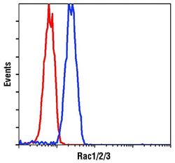
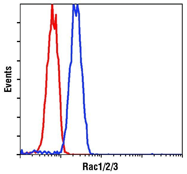
- Flow cytometric analysis of Rac1/2/3 in Jurkat cells using a Rac1/2/3 polyclonal antibody (Product # PA5-17519) (blue) compared to a nonspecific negative control antibody (red).
Supportive validation
- Submitted by
- Invitrogen Antibodies (provider)
- Main image
- Experimental details
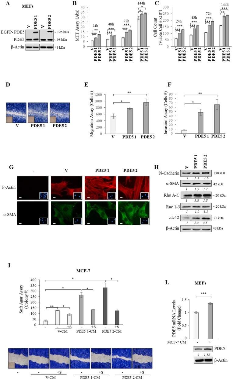
- Figure 4 PDE5 overexpression and fibroblast activation. ( A ) Immunoblotting for PDE5 expression in mouse embryonic fibroblasts (MEFs) stably transfected with pEGFP (enhanced-green-fluorescent-protein) vector (V) and pEGFP-PDE5A expression plasmid (PDE5 1 and PDE5 2). beta-Actin was used as a control for equal loading and transfer. Italicized numbers below blots represent the mean of the band optical density expressed as fold over V for PDE1 and PDE2. ( B ) MTT growth and ( C ) Trypan blue cell count assays in V, PDE5 1, and PDE5 2 stable clones under basal nonstimulated conditions at indicated times. ( D ) Wound healing assay in V, PDE5 1, and PDE5 2 stable clones with images captured at 0 (inset) and 12 h. Pictures are representative of three independent experiments. ( E ) Boyden chamber transmigration and ( F ) invasion assays in V, PDE5 1, and PDE5 2 stable clones under basal nonstimulated conditions. ( G ) Immunofluorescent staining of phalloidin staining of F-actin (stress fibers, red, upper panel) and alpha-SMA (lower panel) and in stable clones. 4',6-Diamidino-2-phenylindole (DAPI) staining was used for nuclei detection (inset). Pictures are representative of three independent experiments. Scale bar = 5 um. ( H ) Immunoblotting for N-cadherin, alpha-SMA, Rho A-C, Rac 1-3, and cdc42 expression levels in V, PDE5 1, and PDE5 2 stable clones. beta-Actin was used as a control for equal loading and transfer. Italicized numbers below blots represent the mean of the band op