 Explore
 Validate
 Learn
Learn Western blot
Antibody data
- Antibody Data
- Antigen structure
- References [1]
- Comments [0]
- Validations
- Western blot [3]
- Immunohistochemistry [1]
Submit
Validation data
Reference
Comment
Report error
- Product number
- PA5-48512 - Provider product page

- Provider
- Invitrogen Antibodies
- Product name
- PQLC2 Polyclonal Antibody
- Antibody type
- Polyclonal
- Antigen
- Synthetic peptide
- Reactivity
- Human, Mouse
- Host
- Rabbit
- Isotype
- IgG
- Vial size
- 400 µL
- Concentration
- 0.4 mg/mL
- Storage
- Store at 4°C short term. For long term storage, store at -20°C, avoiding freeze/thaw cycles.
Submitted references Macrophage Metabolism of Apoptotic Cell-Derived Arginine Promotes Continual Efferocytosis and Resolution of Injury.
Yurdagul A Jr, Subramanian M, Wang X, Crown SB, Ilkayeva OR, Darville L, Kolluru GK, Rymond CC, Gerlach BD, Zheng Z, Kuriakose G, Kevil CG, Koomen JM, Cleveland JL, Muoio DM, Tabas I
Cell metabolism 2020 Mar 3;31(3):518-533.e10
Cell metabolism 2020 Mar 3;31(3):518-533.e10
No comments: Submit comment
Supportive validation
- Submitted by
- Invitrogen Antibodies (provider)
- Main image
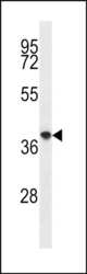
- Experimental details
- Western blot analysis of PQLC2 in in mouse spleen tissue lysates (35µg/lane). Samples were probed with a PQLC2 Antibody (C-term) (Product # PA5-48512).

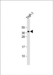
- Submitted by
- Invitrogen Antibodies (provider)
- Main image

- Experimental details
- Western blot analysis of PQLC2. Anti-PQLC2 Antibody (C-term) (Product # PA5-48512) at a 1:1000 dilution + THP-1 whole cell lysate.

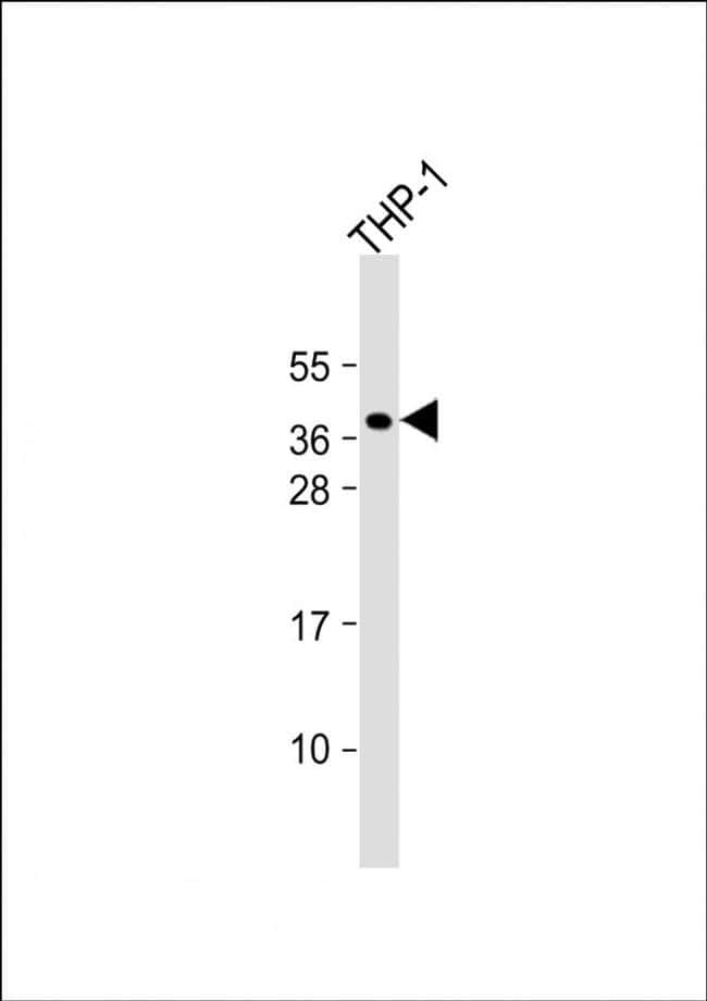
- Submitted by
- Invitrogen Antibodies (provider)
- Main image
- Experimental details
- Western blot analysis of PQLC2. Anti-PQLC2 Antibody (C-term) (Product # PA5-48512) at a 1:1000 dilution + THP-1 whole cell lysate.
Supportive validation
- Submitted by
- Invitrogen Antibodies (provider)
- Main image

- Experimental details
- Immunohistochemical analysis of PQLC2 in formalin fixed and paraffin embedded human testis tissue. Samples stained using a PQLC2 Antibody (C-term) (Product # PA5-48512), followed by peroxidase conjugation of the secondary antibody and DAB staining.
