Explore
 Validate
 Learn
Learn Western blot
 ELISA
ELISA Immunocytochemistry
Antibody data
- Antibody Data
- Antigen structure
- References [0]
- Comments [0]
- Validations
- Immunocytochemistry [1]
Submit
Validation data
Reference
Comment
Report error
- Product number
- MA1-10808 - Provider product page

- Provider
- Invitrogen Antibodies
- Product name
- Hemoglobin Monoclonal Antibody (HB11-231.2)
- Antibody type
- Monoclonal
- Antigen
- Purifed from natural sources
- Description
- MA1-10808 detects Hemoglobin from human samples. This antibody does not detect bovine, chicken, equine, ovine, or porcine Hemoglobin. MA1-10808 has been successfully used in ELISA and Western blot applications. The MA1-10808 immunogen is purified human hemoglobin.
- Reactivity
- Human
- Host
- Mouse
- Isotype
- IgG
- Antibody clone number
- HB11-231.2
- Vial size
- 500 μg
- Concentration
- 0.9 mg/mL
- Storage
- -20°C, Avoid Freeze/Thaw Cycles
No comments: Submit comment
Supportive validation
- Submitted by
- Invitrogen Antibodies (provider)
- Main image
- Experimental details
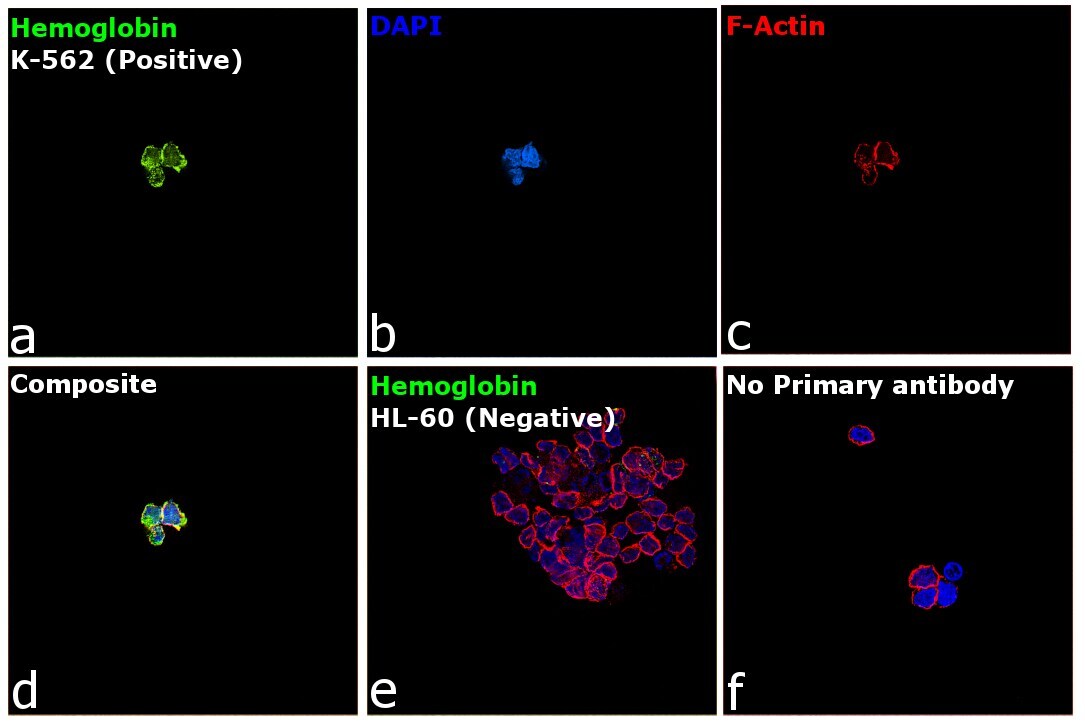
- Immunofluorescence analysis of Hemoglobin was performed using K-562 cells. The cells were fixed with 4% paraformaldehyde for 10 minutes, permeabilized with 0.1% Triton™ X-100 for 15 minutes, and blocked with 2% BSA for 45 minutes at room temperature. The cells were labeled with Hemoglobin Monoclonal Antibody (HB11-231.2) (Product # MA1-10808) at 1:100 dilution in 0.1% BSA, incubated at 4 degree celsius overnight and then labeled with Donkey anti-Mouse IgG (H+L) Highly Cross-Adsorbed Secondary Antibody, Alexa Fluor Plus 488 (Product # A32766), (1:2000 dilution), for 45 minutes at room temperature (Panel a: Green). Nuclei (Panel b: Blue) were stained with ProLong™ Diamond Antifade Mountant with DAPI (Product # P36962). F-actin (Panel c: Red) was stained with Rhodamine Phalloidin (Product # R415, 1:300). Panel d represents the merged image showing Cytoplasmic localization. Panel e represents HL-60 cells having no expression of Hemoglobin. Panel f represents control cells with no primary antibody to assess background. The images were captured at 60X magnification.