Explore
 Validate
 Learn
Learn Western blot
 ELISA
ELISAAntibody data
- Antibody Data
- Antigen structure
- References [0]
- Comments [0]
- Validations
- Western blot [2]
- Immunohistochemistry [1]
Submit
Validation data
Reference
Comment
Report error
- Product number
- GTX23627 - Provider product page

- Provider
- GeneTex
- Proper citation
- GeneTex Cat#GTX23627, RRID:AB_368898
- Product name
- LDB2 antibody
- Antibody type
- Polyclonal
- Reactivity
- Human, Mouse, Rat, Canine, Chicken/Avian
- Host
- Rabbit
No comments: Submit comment
Supportive validation
- Submitted by
- GeneTex (provider)
- Main image
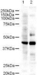
- Experimental details
- Western blot using GeneTex's Affinity Purified anti-LDB2 antibody shows detection of a 43-kDa band corresponding to LDB2 in a lysates prepared from human kidney (lane 1) and mouse spleen (lane 2) tissues. Approximately 18 µg of lysate was run on a SDS-PAGE and transferred onto nitrocellulose followed by reaction with a 1:500 dilution of anti-LDB2 antibody. Detection occurred using a 1:5,000 dilution of HRP-labeled Goat anti-Rabbit IgG for 1 hour at room temperature. A chemiluminescence system was used for signal detection (Roche) using a 1 min exposure time.
- Validation comment
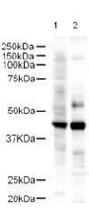
- WB

- Submitted by
- GeneTex (provider)
- Main image

- Experimental details
- Western blot using GeneTex's Affinity Purified anti-LDB2 antibody shows detection of a 43-kDa band corresponding to LDB2 in a lysates prepared from human kidney (lane 1) and mouse spleen (lane 2) tissues. Approximately 18 ?g of lysate was run on a SDS-PAGE and transferred onto nitrocellulose followed by reaction with a 1:500 dilution of anti-LDB2 antibody. Detection occurred using a 1:5,000 dilution of HRP-labeled Goat anti-Rabbit IgG for 1 hour at room temperature. A chemiluminescence system was used for signal detection (Roche) using a 1 min exposure time.
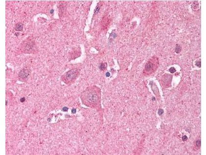
Supportive validation
- Submitted by
- GeneTex (provider)
- Main image
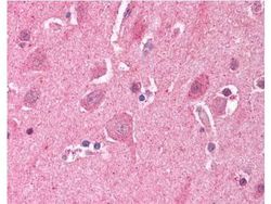
- Experimental details
- GeneTex's Affinity Purified anti-LDB2 (Clim1) antibody was used at a 5 ?g/ml to detect LDB2 in human brain cortex tissue. The image shows the localization of antibody as the precipitated red signal, with a hematoxylin purple nuclear counter stain. Tissue was formalin-fixed and paraffin embedded.