Explore
 Validate
 Learn
Learn Western blot
Antibody data
- Antibody Data
- Antigen structure
- References [0]
- Comments [0]
- Validations
- Western blot [2]
- Immunocytochemistry [2]
Submit
Validation data
Reference
Comment
Report error
- Product number
- 710400 - Provider product page

- Provider
- Invitrogen Antibodies
- Product name
- PI3K p85/p55 Recombinant Polyclonal Antibody (6HCLC)
- Antibody type
- Polyclonal
- Antigen
- Synthetic peptide
- Reactivity
- Human, Mouse
- Host
- Rabbit
- Isotype
- IgG
- Antibody clone number
- 6HCLC
- Vial size
- 100 µg
- Concentration
- 0.5 mg/mL
- Storage
- Store at 4°C short term. For long term storage, store at -20°C, avoiding freeze/thaw cycles.
No comments: Submit comment
Supportive validation
- Submitted by
- Invitrogen Antibodies (provider)
- Main image

- Experimental details
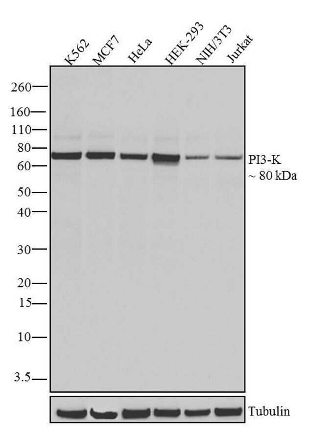
- Western blot analysis of PI3K was performed by loading 20 µg of K562 (lane1), MCF7 (lane2), HeLa (lane3), HEK-293 (lane4), NIH/3T3 (lane5) and Jurkat (lane6) cell lysates using Novex®NuPAGE®4-12 % Bis-Tris gel (Product # NP0321BOX), XCell SureLock Electrophoresis System (Product # EI0002), Novex® Sharp Pre-Stained Protein Standard (Product # LC5800), and iBlot® Dry Blotting System (Product # IB21001). Proteins were transferred to a nitrocellulose membrane and blocked with 5 % skim milk for 1 hour at room temperature. PI3K was detected at ~80 kDa using PI3K Recombinant Rabbit Polyclonal Antibody (Product # 710400) at 0.5 µg-1 µg/mL in 2.5 % skim milk at 4°C overnight on a rocking platform. Goat anti-Rabbit IgG-HRP Secondary Antibody (Product # G-21234) at 1:5000 dilution was used and chemiluminescent detection was performed using Pierce™ ECL Western blotting Substrate (Product # 32106).
- Submitted by
- Invitrogen Antibodies (provider)
- Main image

- Experimental details
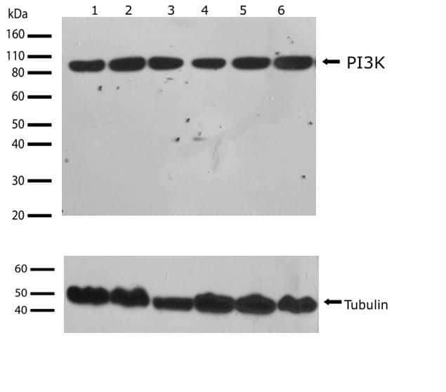
- Western blot analysis of PI3 kinase p85 in whole extracts of Jurkat, NIH-3T3, HEK293, HeLa, MCF-7, and K562 lysate (lanes 1-5 respectively) using a PI3 kinase p85 Recombinant Rabbit Polyclonal Antibody (Product # 710400) at a dilution of 2 µg/mL. Tubulin was used as a loading control and detected with an Anti-tubulin antibody. Samples were detected using chemiluminescence (ECL). Results show a band at ~85kDa.
Supportive validation
- Submitted by
- Invitrogen Antibodies (provider)
- Main image
- Experimental details
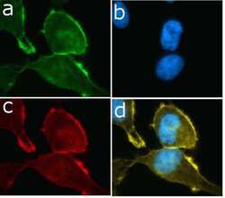
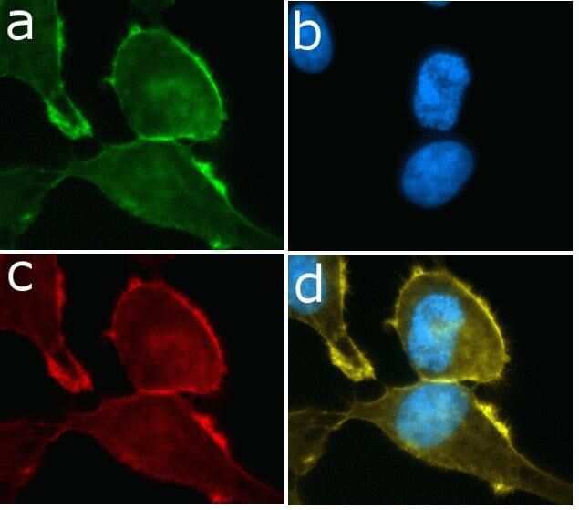
- Immunofluorescent analysis of PI3 kinase p85 in HeLa cells using a PI3 kinase p85 Recombinant Rabbit Polyclonal Antibody (Product # 710400) followed by detection using an Alexa Fluor 488-conjugated Goat anti-Rabbit secondary antibody (green) (Image A). Nuclei were stained using DAPI (Image B) and actin stained with Alexa Fluor 594 phalloidin (red) (image C). Image D is a composite image showing cytoplasmic localization of PI3K.
- Submitted by
- Invitrogen Antibodies (provider)
- Main image
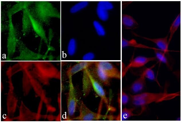
- Experimental details
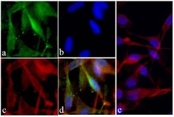
- Immunofluorescent analysis of PI3K was done on 70% confluent log phase U-87 MG cells. The cells were fixed with 4% paraformaldehyde for 15 minutes; permeabilized with 0.25% Triton X-100 for 10 minutes followed by blocking with 5% BSA for 1 hour at room temperature. The cells were incubated with PI3K Recombinant Rabbit Polyclonal Antibody (Product # 710400) at 2 µg-4 µg in 1% BSA and incubated for 3 hours at room temperature and then labeled with Alexa Fluor® 488 Goat anti-Rabbit IgG Secondary Antibody (Product # A-11008) at a dilution of 1:400 for 30 minutes at room temperature (Panel a: green). Nuclei (Panel b: blue) were stained with SlowFade® Gold Antifade Mountant with DAPI (Product # S36938). F-actin (Panel c: red) was stained with Alexa Fluor® 594 Phalloidin (Product # A12381). Panel d is a merged image showing cytoplasmic and membrane localization of PI3K. Panel e shows no primary antibody. The images were captured at 20X magnification.