Explore
 Validate
 Learn
Learn Western blot
Antibody data
- Antibody Data
- Antigen structure
- References [0]
- Comments [0]
- Validations
- Western blot [6]
- Immunohistochemistry [3]
Submit
Validation data
Reference
Comment
Report error
- Product number
- PA5-27987 - Provider product page

- Provider
- Invitrogen Antibodies
- Product name
- Arginase 2 Polyclonal Antibody
- Antibody type
- Polyclonal
- Antigen
- Recombinant protein fragment
- Description
- Recommended positive controls: 293T, NT2D1, 293T cytoplasm extract, mouse kidney. Predicted reactivity: Mouse (88%), Rat (87%), Dog (85%), Rabbit (90%), Chicken (81%), Rhesus Monkey (99%), Bovine (93%). Store product as a concentrated solution. Centrifuge briefly prior to opening the vial.
- Reactivity
- Human, Mouse, Rat
- Host
- Rabbit
- Isotype
- IgG
- Vial size
- 100 µL
- Concentration
- 0.32 mg/mL
- Storage
- Store at 4°C short term. For long term storage, store at -20°C, avoiding freeze/thaw cycles.
No comments: Submit comment
Supportive validation
- Submitted by
- Invitrogen Antibodies (provider)
- Main image
- Experimental details
- Western blot analysis of Arginase II using 30 µg NT2D1 whole cell lysate. Samples were loaded onto a 10% SDS-PAGE gel and probed with an Arginase II polyclonal antibody (Product # PA5-27987) at a dilution of 1:1000.

- Submitted by
- Invitrogen Antibodies (provider)
- Main image
- Experimental details
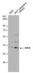
- Western Blot analysis of Arginase 2 was performed by separating 30 µg of 293T whole cell and cytoplasm extracts by 10% SDS-PAGE. Proteins were transferred to a membrane and probed with a Arginase 2 Polyclonal Antibody (Product # PA5-27987) at a dilution of 1:1000. The HRP-conjugated anti-rabbit IgG antibody was used to detect the primary antibody.

- Submitted by
- Invitrogen Antibodies (provider)
- Main image

- Experimental details
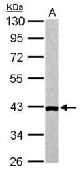
- Western Blot analysis of Arginase 2 was performed by separating 30 µg of whole cell extract by 10% SDS-PAGE. Proteins were transferred to a membrane and probed with a Arginase 2 Polyclonal Antibody (Product # PA5-27987) at a dilution of 1:1000.

- Submitted by
- Invitrogen Antibodies (provider)
- Main image
- Experimental details
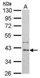
- Arginase II antibody detects ARG2 protein by western blot analysis. A. 50 µg mouse kidney lysate/extract.10% SDS-PAGE. Arginase II antibody Arginase 2 Polyclonal Antibody (Product # PA5-27987) dilution: 1:1,000. The HRP-conjugated anti-rabbit IgG antibody was used to detect the primary antibody.

- Submitted by
- Invitrogen Antibodies (provider)
- Main image
- Experimental details
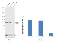
- Knockdown of Arginase 2 was achieved by transfecting LNCaP with Arginase 2 specific siRNA (Silencer® select Product # s1571). Western blot analysis (Fig. a) was performed using Whole cell extracts from the Arginase 2 knockdown cells (lane 3), non-specific scrambled siRNA transfected cells (lane 2) and untransfected cells (lane 1). The blot was probed with Arginase 2 Polyclonal Antibody (Product # PA5-27987, 1:2000 dilution) and Goat anti-Rabbit IgG (H+L) Superclonal™ Recombinant Secondary Antibody, HRP (Product # A27036, 0.25µg/ml, 1:4000 dilution). Densitometric analysis of this western blot is shown in histogram (Fig. b). Decrease in signal upon siRNA mediated knock down confirms that antibody is specific to Arginase 2.

- Submitted by
- Invitrogen Antibodies (provider)
- Main image
- Experimental details
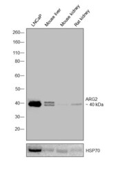
- Western blot was performed using Anti-Arginase 2 Polyclonal Antibody (Product # PA5-27987) and a 40 kDa band corresponding to Arginase 2 was observed across cell line and tissues tested. Whole cell extracts (40 µg lysate) of LNCaP (Lane 1), Mouse liver (Lane 2), Mouse kidney (Lane 3), and Rat kidney (Lane 4) were electrophoresed using Novex® NuPAGE® 4-12 % Bis-Tris gel (Product # NP0321BOX). Resolved proteins were then transferred onto a nitrocellulose membrane (Product # IB23001) by iBlot® 2 Dry Blotting System (Product # IB21001). The blot was probed with the primary antibody (1:1000 dilution) and detected by chemiluminescence with Goat anti-Rabbit IgG (H+L), Superclonal™ Recombinant Secondary Antibody, HRP conjugate (Product # A27036, 1:4000 dilution) using the iBright FL 1000 (Product # A32752). Chemiluminescent detection was performed using Novex® ECL Chemiluminescent Substrate Reagent Kit (Product # WP20005).

Supportive validation
- Submitted by
- Invitrogen Antibodies (provider)
- Main image
- Experimental details
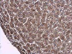
- Immunohistochemistry (Paraffin) analysis of Arginase 2 was performed in paraffin-embedded rat liver tissue using Arginase 2 Polyclonal Antibody (Product # PA5-27987) at a dilution of 1:500.

- Submitted by
- Invitrogen Antibodies (provider)
- Main image
- Experimental details
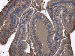
- Immunohistochemistry (Paraffin) analysis of Arginase 2 was performed in paraffin-embedded mouse duodenum tissue using Arginase 2 Polyclonal Antibody (Product # PA5-27987) at a dilution of 1:500.

- Submitted by
- Invitrogen Antibodies (provider)
- Main image
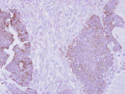
- Experimental details
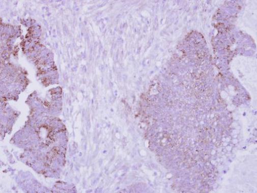
- Immunohistochemical analysis of paraffin-embedded human colon carcinoma, using Arginase II (Product # PA5-27987) antibody at 1:250 dilution. Antigen Retrieval: EDTA based buffer, pH 8.0, 15 min.