Explore
 Validate
 Learn
Learn Western blot
Antibody data
- Antibody Data
- Antigen structure
- References [0]
- Comments [0]
- Validations
- Western blot [1]
- Immunocytochemistry [2]
- Immunohistochemistry [4]
Submit
Validation data
Reference
Comment
Report error
- Product number
- 103-M80 - Provider product page

- Provider
- ReliaTech GmbH
- Product name
- Emilin-1
- Antibody type
- Monoclonal
- Antigen
- Recombinant mouse Emilin-1
- Description
- antibody purified from hybridoma supernatant
- Reactivity
- Mouse
- Host
- Rat
- Antibody clone number
- (#C11A8)
- Vial size
- 100 µl
- Storage
- Store lyophilized at 2-8°C for 6 months or at -20°C long term. After reconstitution store the antibody undiluted at 2-8°C for one month or (in aliquots) at -20°C long term. Avoid repeated freezing and thawing.
- Handling
- Restore in sterile water to a concentration of 0.1-1.0 mg/ml. The antibody solution should be gently mixed before use.
No comments: Submit comment
Supportive validation
- Submitted by
- ReliaTech GmbH (provider)
- Main image
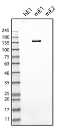
- Experimental details
- Western blot analysis on recombinant protein obtained from supernatant of cells expressing human EMILIN1 (hE1), mouse EMILIN1 (mE1), mouse EMILIN2 (mE2).

Supportive validation
- Submitted by
- ReliaTech GmbH (provider)
- Main image
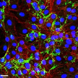
- Experimental details
- Positive staining of EMILIN1 (green) in mouse fibroblasts (NIH 3T3). Actin cytoskeleton is stained in red, nuclei in blue. Scale bar 28 μm.
- Sample type
- mouse fibroblasts (NIH 3T3)

- Submitted by
- ReliaTech GmbH (provider)
- Main image
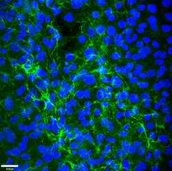
- Experimental details
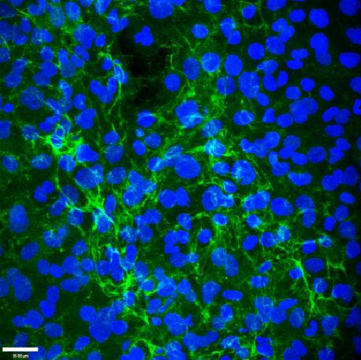
- Positive staining of EMILIN1 (green) in mouse lymphatic endothelial cells (LAEC). Nuclei are stained in blue. Scale bar 38 μm.
- Sample type
- mouse lymphatic endothelial cells (LAEC)
Supportive validation
- Submitted by
- ReliaTech GmbH (provider)
- Main image
- Experimental details
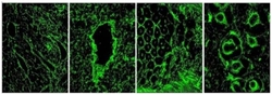
- Cryostat sections of normal mouse tissues stained with anti-Emilin-1 antibodies. In all mouse tissues and organs examined, Emilin-1 was uniformly distributed in the stroma. In the skin, Emilin-1 staining colocalizes with LYVE-1-positive lymphatic vessels surrounding hair follicles. In the small intestine, it colocalizes with LYVE-1-positive lacteals and submucosal lymphatic vessels. At higher magnification, in the lung and lymph nodes, it is more evident that Emilin-1 is distributed at the abluminal surfaces of LECs . In the lymph node, Emilin-1-positive fibers connecting LECs to the surrounding ECM are evident.
- Sample type
- Mouse tissue

- Submitted by
- ReliaTech GmbH (provider)
- Main image

- Experimental details
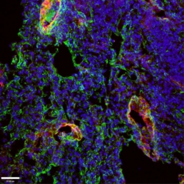
- Positive staining for EMILIN1 (green) in mouse lung tissue is both detected in lung parenchymal fibers and associated with lymphatic vessel structures. Lyve-1 (red) is used as lymphatic vessel marker. Blue, nuclei. Scale bar: 37.00 μm
- Submitted by
- ReliaTech GmbH (provider)
- Main image
- Experimental details
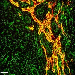
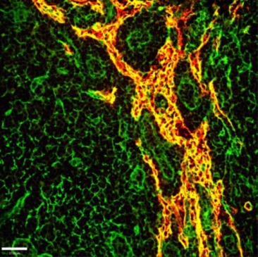
- Positive staining for EMILIN1 (green) in mouse lymph node. In red, lymphatic vessels positive for Lyve-1. Yellow represents the close association of EMILIN1 with lymphatic vessel structures. Scale bar: 37.00 μm
- Sample type
- mouse lymph node
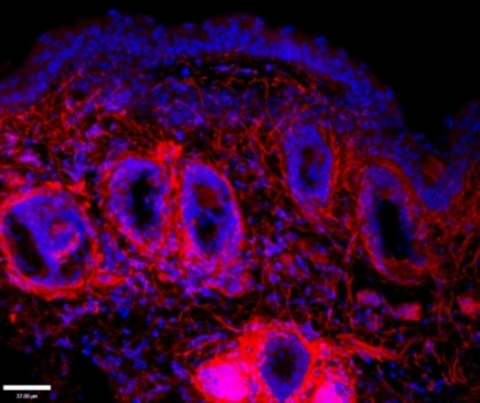
- Submitted by
- ReliaTech GmbH (provider)
- Main image
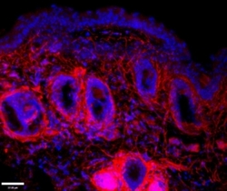
- Experimental details
- Positive staining for EMILIN1 (red) in mouse skin. Blue, nuclei; Scale bar: 37.00 μm
- Sample type
- mouse skin