Explore
 Validate
 Learn
Learn Immunocytochemistry
 Immunohistochemistry
Antibody data
- Antibody Data
- Antigen structure
- References [1]
- Comments [0]
- Validations
- Immunohistochemistry [1]
- Flow cytometry [2]
Submit
Validation data
Reference
Comment
Report error
- Product number
- MAB8164 - Provider product page

- Provider
- Novus Biologicals
- Product name
- Mouse Monoclonal TM4SF1/L6 Antibody
- Antibody type
- Monoclonal
- Description
- Protein A or G purified from hybridoma culture supernatant. Detects human TM4SF1/L6 in ELISA. In Flow Cytometry, it stains HEK293 cells transfected with human TM4SF1/L6, and does not stain HEK293 cells transfected with irrelevant human cell surface marker.
- Reactivity
- Human
- Host
- Mouse
- Conjugate
- Unconjugated
- Isotype
- IgG
- Vial size
- 100 ug
- Concentration
- LYOPH
- Storage
- Use a manual defrost freezer and avoid repeated freeze-thaw cycles. 12 months from date of receipt, -20 to -70 degreesC as supplied. 1 month, 2 to 8 degreesC under sterile conditions after reconstitution. 6 months, -20 to -70 degreesC under sterile conditions after reconstitution.
Submitted references Regeneration of the lung alveolus by an evolutionarily conserved epithelial progenitor.
Zacharias WJ, Frank DB, Zepp JA, Morley MP, Alkhaleel FA, Kong J, Zhou S, Cantu E, Morrisey EE
Nature 2018 Mar 8;555(7695):251-255
Nature 2018 Mar 8;555(7695):251-255
No comments: Submit comment
Supportive validation
- Submitted by
- Novus Biologicals (provider)
- Main image
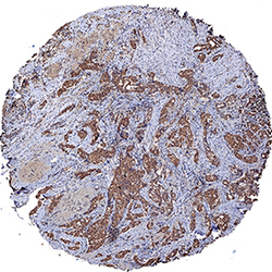
- Experimental details
- TM4SF1/L6 in Human Breast Cancer Tissue. TM4SF1/L6 was detected in immersion fixed paraffin-embedded sections of human breast cancer tissue using Mouse Anti-Human TM4SF1/L6 Monoclonal Antibody (Catalog # MAB8164) at 5 µg/mL for 1 hour at room temperature followed by incubation with the Anti-Mouse IgG VisUCyte™ HRP Polymer Antibody (Catalog # VC001). Tissue was stained using DAB (brown) and counterstained with hematoxylin (blue). Specific staining was localized to cytoplasm in cancer cells. View our protocol for IHC Staining with VisUCyte HRP Polymer Detection Reagents.

Supportive validation
- Submitted by
- Novus Biologicals (provider)
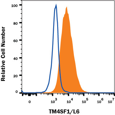
- Main image
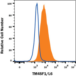
- Experimental details
- Detection of TM4SF1/L6 in A549 Human Cell Line by Flow Cytometry. A549 human lung carcinoma cell line was stained with Mouse Anti-Human TM4SF1/L6 Monoclonal Antibody (Catalog # MAB8164, filled histogram) or isotype control antibody (Catalog # MAB002, open histogram), followed by Allophycocyanin-conjugated Anti-Mouse IgG Secondary Antibody (Catalog # F0101B). View our protocol for Staining Membrane-associated Proteins.
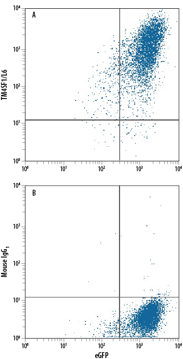
- Submitted by
- Novus Biologicals (provider)
- Main image
- Experimental details
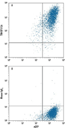
- Detection of TM4SF1/L6 in HEK293 Human Cell Line Transfected with Human TM4SF1/L6 and eGFP by Flow Cytometry. HEK293 human embryonic kidney cell line transfected with human TM4SF1/L6 and eGFP was stained with either (A) Mouse Anti-Human TM4SF1/L6 Monoclonal Antibody (Catalog # MAB8164) or (B) Mouse IgG1 Isotype Control (Catalog # MAB002) followed by Phycoerythrin-conjugated Anti-Mouse IgG Secondary Antibody (Catalog # F0102B). View our protocol for Staining Membrane-associated Proteins.