Explore
 Validate
 Learn
LearnAGC-017-200UL
antibody from Invitrogen Antibodies
Targeting: GRM7
GLUR7, GPRC1G, mGlu7, MGLUR7, PPP1R87
 Western blot
Antibody data
- Antibody Data
- Antigen structure
- References [0]
- Comments [0]
- Validations
- Western blot [2]
- Immunocytochemistry [1]
- Immunohistochemistry [1]
- Flow cytometry [1]
Submit
Validation data
Reference
Comment
Report error
- Product number
- AGC-017-200UL - Provider product page

- Provider
- Invitrogen Antibodies
- Product name
- mGluR7 (extracellular) Polyclonal Antibody
- Antibody type
- Polyclonal
- Antigen
- Other
- Reactivity
- Human, Mouse, Rat
- Host
- Rabbit
- Isotype
- IgG
- Vial size
- 200 µL
- Concentration
- 0.8 mg/mL
- Storage
- -20° C, Avoid Freeze/Thaw Cycles
No comments: Submit comment
Supportive validation
- Submitted by
- Invitrogen Antibodies (provider)
- Main image
- Experimental details
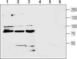
- Western blot analysis of mouse (lanes 1 and 4) and rat (lanes 2 and 5) brain membranes and human CCF-STTGI astrocytoma (lanes 3 and 6) cell line lysate (1:200): - 1-3. Anti-mGluR7 (extracellular) Antibody (#AGC-017), (1:200).4-6. Anti-mGluR7 (extracellular) Antibody , preincubated with mGluR7 (extracellular) Blocking Peptide (#BLP-GC017).

- Submitted by
- Invitrogen Antibodies (provider)
- Main image

- Experimental details
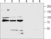
- Western blot analysis of mouse (lanes 1 and 4) and rat (lanes 2 and 5) brain membranes and human CCF-STTGI astrocytoma (lanes 3 and 6) cell line lysate (1:200): - 1-3. Anti-mGluR7 (extracellular) Antibody (#AGC-017), (1:200).4-6. Anti-mGluR7 (extracellular) Antibody , preincubated with mGluR7 (extracellular) Blocking Peptide (#BLP-GC017).
Supportive validation
- Submitted by
- Invitrogen Antibodies (provider)
- Main image
- Experimental details
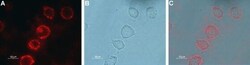
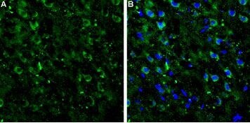
- Expression of mGluR7 in rat PC12 cells - Cell surface detection of mGluR7 in live intact rat pheochromocytoma PC12 cells. A. Cells were stained with Anti-mGluR7 (extracellular) Antibody (#AGC-017), (1:100), followed by goat Anti-rabbit-AlexaFluor-594 secondary Antibody (red). B. Live view of the cells. C. Merge of the two pictures.

Supportive validation
- Submitted by
- Invitrogen Antibodies (provider)
- Main image
- Experimental details
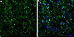
- Expression of mGluR7 in rat neocortex - Immunohistochemical staining of rat neocortex frozen sections using Anti-mGluR7 (extracellular) Antibody (#AGC-017).A.mGluR7 staining (green) appears in several neocortical neurons. B. Merge image showing mGluR7 staining together with cell nuclei (blue).
Supportive validation
- Submitted by
- Invitrogen Antibodies (provider)
- Main image
- Experimental details
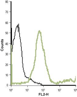
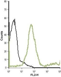
- Cell surface detection of mGluR7 in live intact human T cell leukemia (Jurkat) cell line: - (black) Unstained cells + goat- Anti-rabbit-PE. (green) Cells + Anti-mGluR7 (extracellular) Antibody (#AGC-017), (1:20) + goat- Anti-rabbit-PE.