 Explore
 Validate
 Learn
Learn Western blot
Antibody data
- Antibody Data
- Antigen structure
- References [1]
- Comments [0]
- Validations
- Western blot [2]
- Immunohistochemistry [1]
Submit
Validation data
Reference
Comment
Report error
- Product number
- MAB4499 - Provider product page

- Provider
- Novus Biologicals
- Product name
- Mouse Monoclonal PTK7/CCK4 Antibody
- Antibody type
- Monoclonal
- Description
- Protein A or G purified from hybridoma culture supernatant. Detects human and rat PTK7/CCK4 in Western blots.
- Reactivity
- Human, Rat
- Host
- Mouse
- Isotype
- IgG
- Vial size
- 100 ug
- Concentration
- LYOPH
- Storage
- Use a manual defrost freezer and avoid repeated freeze-thaw cycles. 12 months from date of receipt, -20 to -70 degreesC as supplied. 1 month, 2 to 8 degreesC under sterile conditions after reconstitution. 6 months, -20 to -70 degreesC under sterile conditions after reconstitution.
Submitted references The Increased PTK7 Expression Is a Malignant Factor in Cervical Cancer.
Sun JJ, Li HL, Guo SJ, Ma H, Liu SJ, Liu D, Xue FX
Disease markers 2019;2019:5380197
Disease markers 2019;2019:5380197
No comments: Submit comment
Supportive validation
- Submitted by
- Novus Biologicals (provider)
- Main image

- Experimental details
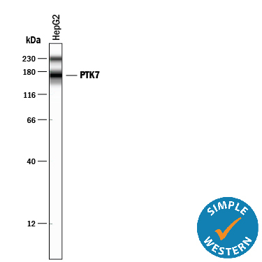
- Detection of Human PTK7/CCK4 by Simple WesternTM. Simple Western lane view shows lysates of HepG2 human hepatocellular carcinoma cell line, loaded at 0.2 mg/mL. A specific band was detected for PTK7/CCK4 at approximately 168 kDa (as indicated) using 20 µg/mL of Mouse Anti-Human/Rat PTK7/CCK4 Monoclonal Antibody (Catalog # MAB4499). This experiment was conducted under reducing conditions and using the 12-230 kDa separation system. Non-specific interaction with the 230 kDa Simple Western standard may be seen with this antibody.
- Submitted by
- Novus Biologicals (provider)
- Main image
- Experimental details
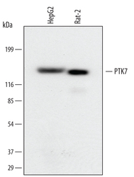
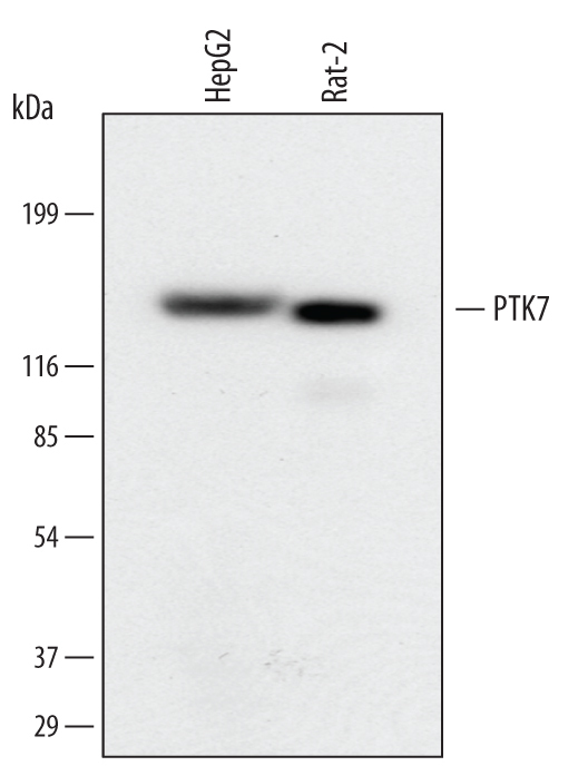
- Detection of Human and Rat PTK7/CCK4 by Western Blot. Western blot shows lysates of HepG2 human hepatocellular carcinoma cell line and Rat-2 rat embryonic fibroblast cell line. PVDF membrane was probed with 1 µg/mL of Mouse Anti-Human/Rat PTK7/CCK4 Monoclonal Antibody (Catalog # MAB4499) followed by HRP-conjugated Anti-Mouse IgG Secondary Antibody (Catalog # HAF007). A specific band was detected for PTK7/CCK4 at approximately 160 kDa (as indicated). This experiment was conducted under reducing conditions and using Immunoblot Buffer Group 1.
Supportive validation
- Submitted by
- Novus Biologicals (provider)
- Main image
- Experimental details
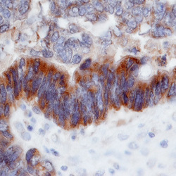
- PTK7/CCK4 in Human Colon Cancer Tissue. PTK7/CCK4 was detected in immersion fixed paraffin-embedded sections of human colon cancer tissue using Mouse Anti-Human/Rat PTK7/CCK4 Monoclonal Antibody (Catalog # MAB4499) at 25 µg/mL overnight at 4 °C. Tissue was stained using the Anti-Mouse HRP-DAB Cell & Tissue Staining Kit (brown; Catalog # CTS002) and counterstained with hematoxylin (blue). Specific labeling was localized to the cytoplasm of epithelial cells. View our protocol for Chromogenic IHC Staining of Paraffin-embedded Tissue Sections.
