Explore
 Validate
 Learn
Learn Western blot
 Immunocytochemistry
Antibody data
- Antibody Data
- Antigen structure
- References [2]
- Comments [0]
- Validations
- Immunocytochemistry [3]
- Immunohistochemistry [1]
Submit
Validation data
Reference
Comment
Report error
- Product number
- MA1-5785 - Provider product page

- Provider
- Invitrogen Antibodies
- Product name
- HEF1 Monoclonal Antibody (2G9)
- Antibody type
- Monoclonal
- Antigen
- Other
- Description
- This antibody detects HEF1 in human, mouse, and rat samples. This antibody does not recognize p130cas. It has not been tested on Sin1. This antibody mostly detects HEF1 localized at the focal adhesion sites.
- Reactivity
- Human, Mouse, Rat
- Host
- Mouse
- Isotype
- IgG
- Antibody clone number
- 2G9
- Vial size
- 100 μL
- Concentration
- 1 mg/mL
- Storage
- Store at 4°C short term. For long term storage, store at -20°C, avoiding freeze/thaw cycles.
Submitted references p75NTR-dependent Rac1 activation requires receptor cleavage and activation of an NRAGE and NEDD9 signaling cascade.
A novel inhibitor of focal adhesion signaling induces caspase-independent cell death in diffuse large B-cell lymphoma.
Zeinieh M, Salehi A, Rajkumar V, Barker PA
Journal of cell science 2015 Feb 1;128(3):447-59
Journal of cell science 2015 Feb 1;128(3):447-59
A novel inhibitor of focal adhesion signaling induces caspase-independent cell death in diffuse large B-cell lymphoma.
Bosch R, Dieguez-Gonzalez R, Céspedes MV, Parreño M, Pavón MÁ, Grañena A, Sierra J, Mangues R, Casanova I
Blood 2011 Oct 20;118(16):4411-20
Blood 2011 Oct 20;118(16):4411-20
No comments: Submit comment
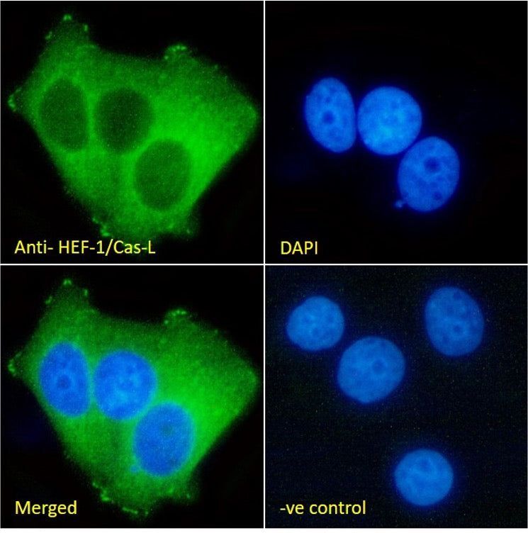
Supportive validation
- Submitted by
- Invitrogen Antibodies (provider)
- Main image
- Experimental details
- Immunocytochemistry analysis of HEF1 in MDA-MB231 cells. Samples were incubated with HEF1 monoclonal antibody (Product # MA1-5785) using a dilution of 1:200. Cells were fixed in 3.8% PFA for 10 minutes, and staining was performed for 1 hour at room temperature.
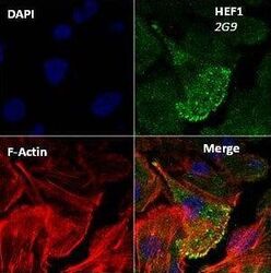
- Submitted by
- Invitrogen Antibodies (provider)
- Main image

- Experimental details
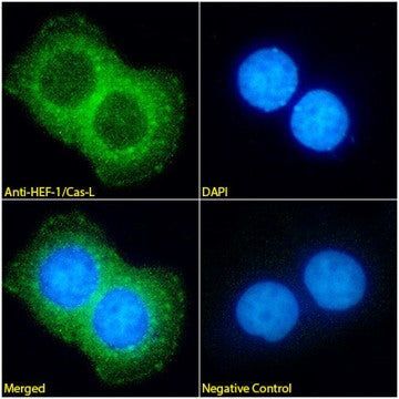
- Immunocytochemistry analysis of HEF1 in paraformaldehyde fixed MCF7 cells, permeabilized with 0.15% Triton. Samples incubated with HEF1 monoclonal antibody (Product # MA1-5785) using a 1:100 dilution for 1hr. Followed by Alexa Fluor® 488 secondary antibody at a 1:1,000 dilution, showing cytoplasmic staining. The nuclear stain is DAPI (blue). Negative control: Mouse IgG1 negative control followed by Alexa Fluor® 488 secondary antibody.
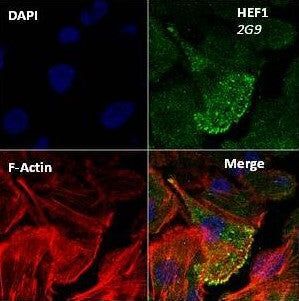
- Submitted by
- Invitrogen Antibodies (provider)
- Main image

- Experimental details
- Immunocytochemistry analysis of Gemin 2 in paraformaldehyde fixed MCF7 cells, permeabilized with 0.15% Triton. Samples were incubated with HEF1 monoclonal antibody (Product # MA1-5785) using a dilution of 1:100 for 1hr. Followed by Alexa Fluor® 488 secondary antibody at a 1:1,000 dilution, showing cytoplasmic staining. The nuclear stain is DAPI (blue). Negative control: Mouse IgG1 negative control followed by Alexa Fluor® 488 secondary antibody.
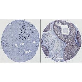
Supportive validation
- Submitted by
- Invitrogen Antibodies (provider)
- Main image

- Experimental details
- Immunohistochemistry analysis of HEF1 in PFA-fixed paraffin embedded normal breast tissue and ductal carcinoma using HEF1 monoclonal antibody (Product # MA1-5785).