Explore
 Validate
 Learn
Learn Western blot
 ELISA
ELISAAntibody data
- Antibody Data
- Antigen structure
- References [3]
- Comments [0]
- Validations
- Western blot [2]
Submit
Validation data
Reference
Comment
Report error
- Product number
- NB100-1699 - Provider product page

- Provider
- Novus Biologicals
- Proper citation
- Novus Cat#NB100-1699, RRID:AB_10000692
- Product name
- Mouse Monoclonal NEDD9/CASL/HEF1 Antibody
- Antibody type
- Monoclonal
- Description
- Protein A purified. Does not recognize p130 Cas. Not tested on Sin1.This mostly detects HEF1 localized at the focal adhesion sites.
- Reactivity
- Human, Mouse
- Host
- Mouse
- Isotype
- IgG
- Vial size
- 0.1 mg
- Concentration
- 1 mg/ml
- Storage
- Store at -20C. Avoid freeze-thaw cycles.
Submitted references Actin polymerization-dependent activation of Cas-L promotes immunological synapse stability.
Knock-in mutation reveals an essential role for focal adhesion kinase activity in blood vessel morphogenesis and cell motility-polarity but not cell proliferation.
The focal adhesion scaffolding protein HEF1 regulates activation of the Aurora-A and Nek2 kinases at the centrosome.
Santos LC, Blair DA, Kumari S, Cammer M, Iskratsch T, Herbin O, Alexandropoulos K, Dustin ML, Sheetz MP
Immunology and cell biology 2016 Nov;94(10):981-993
Immunology and cell biology 2016 Nov;94(10):981-993
Knock-in mutation reveals an essential role for focal adhesion kinase activity in blood vessel morphogenesis and cell motility-polarity but not cell proliferation.
Lim ST, Chen XL, Tomar A, Miller NL, Yoo J, Schlaepfer DD
The Journal of biological chemistry 2010 Jul 9;285(28):21526-36
The Journal of biological chemistry 2010 Jul 9;285(28):21526-36
The focal adhesion scaffolding protein HEF1 regulates activation of the Aurora-A and Nek2 kinases at the centrosome.
Pugacheva EN, Golemis EA
Nature cell biology 2005 Oct;7(10):937-46
Nature cell biology 2005 Oct;7(10):937-46
No comments: Submit comment
Supportive validation
- Submitted by
- Novus Biologicals (provider)
- Main image

- Experimental details
- Western Blot: NEDD9/CASL/HEF1 Antibody (2G9) [NB100-1699] - Detection of a 115 kDa band corresponding to HEF1 in MCF7 lysate (arrowhead). Approximately 35 ig of lysate was loaded for SDS-PAGE followed by transfer onto nitrocellulose and reaction with a 1:1,000 dilution of anti-HEF1 antibody. Detection occurred using a 1:5,000 dilution of IRDye800 conjugated Sh-a-Mouse IgG [H&L] (610-632-002) for 45 min at room temperature (800 nm channel, green). Molecular weight estimation was made by comparison to prestained MW markers (indicated at left). IRDye800 fluorescence image was captured using the OdysseyInfrared Imaging System developed by LI-COR. IRDye is a trademark of LI-COR, Inc. Other detection systems will yield similar results.

- Submitted by
- Novus Biologicals (provider)
- Main image
- Experimental details
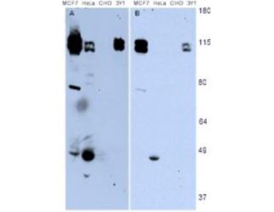
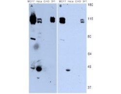
- Western Blot: NEDD9/CASL/HEF1 Antibody (2G9) [NB100-1699] - Detection of endogenous HEF1 present in various cell lines. Panel A shows detection using a 15 min exposure. Panel B is the same blot exposed for 2 min. The doublet represents p105 and p115 staining. The lower MW band represents p55. 3Y1 cells are derived from rat fibroblast cells. Mouse 3T3 cells are also reactive (not shown). To date no staining has been noted on CHO cells.