 Explore
 Validate
 Learn
LearnPA5-28249
antibody from Invitrogen Antibodies
Targeting: OPTN
FIP-2, FIP2, GLC1E, HIP7, HYPL, NRP, TFIIIA-INTP
 Western blot
 Immunocytochemistry
Antibody data
- Antibody Data
- Antigen structure
- References [1]
- Comments [0]
- Validations
- Immunocytochemistry [3]
- Immunoprecipitation [1]
- Immunohistochemistry [1]
- Other assay [2]
Submit
Validation data
Reference
Comment
Report error
- Product number
- PA5-28249 - Provider product page

- Provider
- Invitrogen Antibodies
- Product name
- Optineurin Polyclonal Antibody
- Antibody type
- Polyclonal
- Antigen
- Recombinant full-length protein
- Description
- Recommended positive controls: 293T, A431, H1299, HeLaS3, HepG2, Molt-4, Raji. Predicted reactivity: Mouse (81%), Rat (82%), Pig (84%), Rhesus Monkey (96%), Bovine (86%). Store product as a concentrated solution. Centrifuge briefly prior to opening the vial.
- Reactivity
- Human
- Host
- Rabbit
- Isotype
- IgG
- Vial size
- 100 μL
- Concentration
- 1 mg/mL
- Storage
- Store at 4°C short term. For long term storage, store at -20°C, avoiding freeze/thaw cycles.
Submitted references The optineurin/TIA1 pathway inhibits aberrant stress granule formation and reduces ubiquitinated TDP-43.
Kakihana T, Takahashi M, Katsuragi Y, Yamashita SI, Sango J, Kanki T, Onodera O, Fujii M
iScience 2021 Jul 23;24(7):102733
iScience 2021 Jul 23;24(7):102733
No comments: Submit comment
Supportive validation
- Submitted by
- Invitrogen Antibodies (provider)
- Main image

- Experimental details
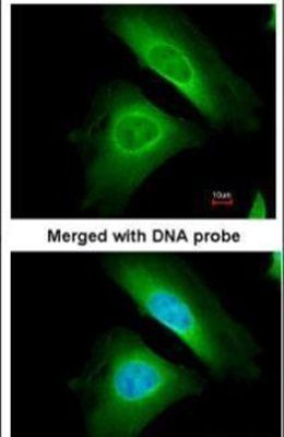
- Immunofluorescent analysis of Optineurin in paraformaldehyde-fixed HeLa cells using an Optineurin polyclonal antibody (Product # PA5-28249) at a 1:200 dilution.
- Submitted by
- Invitrogen Antibodies (provider)
- Main image

- Experimental details
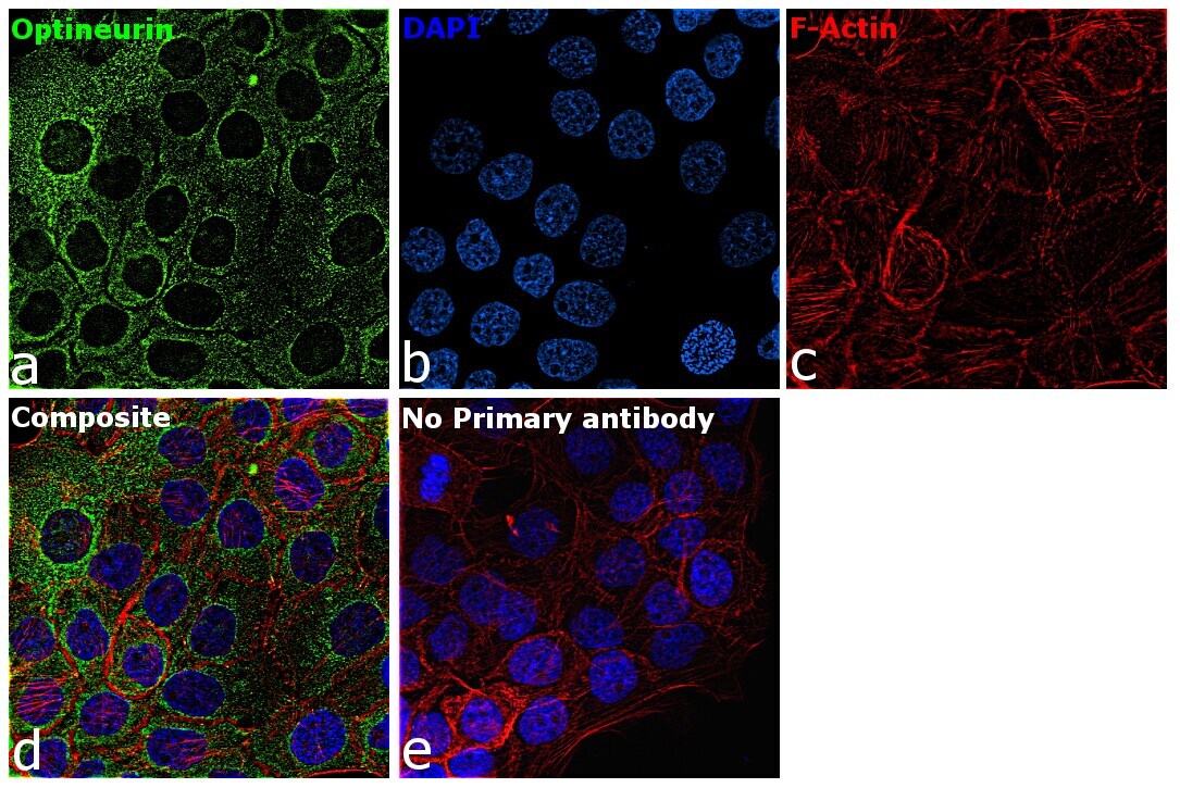
- Immunofluorescence analysis of Optineurin was performed using 70 percent confluent log phase A-431 cells. The cells were fixed with 4% paraformaldehyde for 10 minutes, permeabilized with 0.1% Triton™ X-100 for 15 minutes, and blocked with 2% BSA for 45 minutes at room temperature. The cells were labeled with Optineurin Polyclonal Antibody (Product # PA5-28249) at 1:200 in 0.1% BSA, incubated at 4 degree celsius overnight and then labeled with Goat anti-Rabbit IgG (H+L) Highly Cross-Adsorbed Secondary Antibody, Alexa Fluor Plus 488 (Product # A32731), (1:2000), for 45 minutes at room temperature (Panel a: Green). Nuclei (Panel b:Blue) were stained with ProLong™ Diamond Antifade Mountant with DAPI (Product # P36962). F-actin (Panel c: Red) was stained with Rhodamine Phalloidin (Product # R415, 1:300). Panel d represents the merged image showing cytoplasmic localization. Panel e represents control cells with no primary antibody to assess background. The images were captured at 60X magnification.
- Submitted by
- Invitrogen Antibodies (provider)
- Main image

- Experimental details
- Immunofluorescence analysis of Optineurin was performed using 70 percent confluent log phase A-431 cells. The cells were fixed with 4% paraformaldehyde for 10 minutes, permeabilized with 0.1% Triton™ X-100 for 15 minutes, and blocked with 2% BSA for 45 minutes at room temperature. The cells were labeled with Optineurin Polyclonal Antibody (Product # PA5-28249) at 1:200 in 0.1% BSA, incubated at 4 degree celsius overnight and then labeled with Goat anti-Rabbit IgG (H+L) Highly Cross-Adsorbed Secondary Antibody, Alexa Fluor Plus 488 (Product # A32731), (1:2000), for 45 minutes at room temperature (Panel a: Green). Nuclei (Panel b:Blue) were stained with ProLong™ Diamond Antifade Mountant with DAPI (Product # P36962). F-actin (Panel c: Red) was stained with Rhodamine Phalloidin (Product # R415, 1:300). Panel d represents the merged image showing cytoplasmic localization. Panel e represents control cells with no primary antibody to assess background. The images were captured at 60X magnification.

Supportive validation
- Submitted by
- Invitrogen Antibodies (provider)
- Main image
- Experimental details
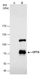
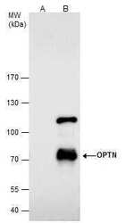
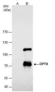
- Optineurin Polyclonal Antibody immunoprecipitates Optineurin protein in IP experiments. IP samples: HepG2 whole cell extract. A. Control with 4 µg of preimmune Rabbit IgG. B. Immunoprecipitation of Optineurin protein by 4 µg Optineurin Polyclonal Antibody (Product # PA5-28249). 7.5 % SDS-PAGE. The immunoprecipitated Optineurin protein was detected by Optineurin Polyclonal Antibody (Product # PA5-28249) diluted at 1:500.
Supportive validation
- Submitted by
- Invitrogen Antibodies (provider)
- Main image
- Experimental details
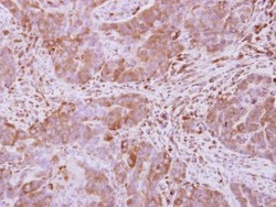
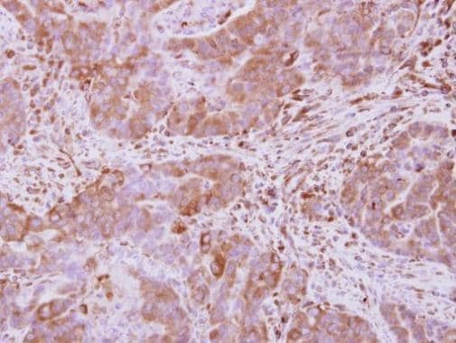
- Immunohistochemical analysis of paraffin-embedded human colon carcinoma, using Optineurin (Product # PA5-28249) antibody at 1:500 dilution. Antigen Retrieval: Citrate buffer, pH 6.0, 15 min.
Supportive validation
- Submitted by
- Invitrogen Antibodies (provider)
- Main image
- Experimental details
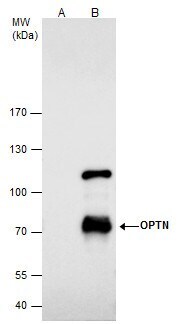
- Optineurin Polyclonal Antibody immunoprecipitates Optineurin protein in IP experiments. IP samples: HepG2 whole cell extract. A. Control with 4 µg of preimmune Rabbit IgG. B. Immunoprecipitation of Optineurin protein by 4 µg Optineurin Polyclonal Antibody (Product # PA5-28249). 7.5 % SDS-PAGE. The immunoprecipitated Optineurin protein was detected by Optineurin Polyclonal Antibody (Product # PA5-28249) diluted at 1:500.
- Submitted by
- Invitrogen Antibodies (provider)
- Main image

- Experimental details
- Optineurin Polyclonal Antibody immunoprecipitates Optineurin protein in IP experiments. IP samples: HepG2 whole cell extract. A. Control with 4 µg of preimmune Rabbit IgG. B. Immunoprecipitation of Optineurin protein by 4 µg Optineurin Polyclonal Antibody (Product # PA5-28249). 7.5 % SDS-PAGE. The immunoprecipitated Optineurin protein was detected by Optineurin Polyclonal Antibody (Product # PA5-28249) diluted at 1:500.
