Explore
 Validate
 Learn
Learn Western blot
 Immunocytochemistry
Antibody data
- Antibody Data
- Antigen structure
- References [7]
- Comments [0]
- Validations
- Immunocytochemistry [7]
- Immunohistochemistry [2]
- Other assay [1]
Submit
Validation data
Reference
Comment
Report error
- Product number
- MA1-611 - Provider product page

- Provider
- Invitrogen Antibodies
- Product name
- Prolactin Receptor Monoclonal Antibody (T6)
- Antibody type
- Monoclonal
- Antigen
- Purifed from natural sources
- Description
- MA1-611 detects prolactin (PRL) receptor in rat and human tissues. This antibody does not cross-react with PRL receptors from other species nor with growth hormone (GH) receptor. MA1-611 has been successfully used in Western blot, IF, immunohistochemistry (4% PFA fixed tissue), and immunocytochemistry procedures. By Western blot, this antibody detects an ~42 kDa protein representing PRL receptor in NB2 cell lysate. Immunohistochemical staining of PRL receptor in NB2 cells with MA1-611 yields a staining pattern consistent with cytoplasmic vesicular staining. MA1-611 has also been used to inhibit the binding of prolactin to prolactin receptor in vitro. The MA1-611 immunogen is purified rat liver PRL receptor. Reconstitute with 100 µL PBS.
- Reactivity
- Human, Rat
- Host
- Mouse
- Isotype
- IgG
- Antibody clone number
- T6
- Vial size
- 100 μg
- Concentration
- 1 mg/mL
- Storage
- -20°C, Avoid Freeze/Thaw Cycles
Submitted references Prolactin secretory rhythm of mated rats induced by a single injection of oxytocin.
Hypothesis paper Brain talks with fat--evidence for a hypothalamic-pituitary-adipose axis?
JAK2 activation and cell proliferation induced by antibody-mediated prolactin receptor dimerization.
Expression of prolactin receptors in murine lymphoid cells in normal and autoimmune situations.
Mutational analysis of the ligand-binding domain of the prolactin receptor.
Identification and functional activity of prolactin receptors in thymic epithelial cells.
Characterization and applications of monoclonal antibodies to the prolactin receptor.
Egli M, Bertram R, Toporikova N, Sellix MT, Blanco W, Freeman ME
American journal of physiology. Endocrinology and metabolism 2006 Mar;290(3):E566-72
American journal of physiology. Endocrinology and metabolism 2006 Mar;290(3):E566-72
Hypothesis paper Brain talks with fat--evidence for a hypothalamic-pituitary-adipose axis?
Schäffler A, Binart N, Schölmerich J, Büchler C
Neuropeptides 2005 Aug;39(4):363-7
Neuropeptides 2005 Aug;39(4):363-7
JAK2 activation and cell proliferation induced by antibody-mediated prolactin receptor dimerization.
Rui H, Lebrun JJ, Kirken RA, Kelly PA, Farrar WL
Endocrinology 1994 Oct;135(4):1299-306
Endocrinology 1994 Oct;135(4):1299-306
Expression of prolactin receptors in murine lymphoid cells in normal and autoimmune situations.
Gagnerault MC, Touraine P, Savino W, Kelly PA, Dardenne M
Journal of immunology (Baltimore, Md. : 1950) 1993 Jun 15;150(12):5673-81
Journal of immunology (Baltimore, Md. : 1950) 1993 Jun 15;150(12):5673-81
Mutational analysis of the ligand-binding domain of the prolactin receptor.
Rozakis-Adcock M, Kelly PA
The Journal of biological chemistry 1991 Sep 5;266(25):16472-7
The Journal of biological chemistry 1991 Sep 5;266(25):16472-7
Identification and functional activity of prolactin receptors in thymic epithelial cells.
Dardenne M, Kelly PA, Bach JF, Savino W
Proceedings of the National Academy of Sciences of the United States of America 1991 Nov 1;88(21):9700-4
Proceedings of the National Academy of Sciences of the United States of America 1991 Nov 1;88(21):9700-4
Characterization and applications of monoclonal antibodies to the prolactin receptor.
Okamura H, Zachwieja J, Raguet S, Kelly PA
Endocrinology 1989 May;124(5):2499-508
Endocrinology 1989 May;124(5):2499-508
No comments: Submit comment
Supportive validation
- Submitted by
- Invitrogen Antibodies (provider)
- Main image
- Experimental details
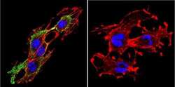
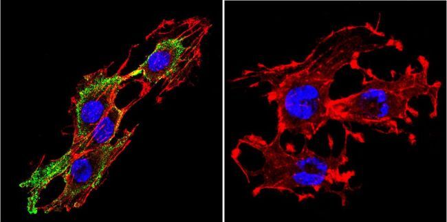
- Immunofluorescent analysis of Prolactin Receptor in C6 Cells. Cells were grown on chamber slides and fixed with formaldehyde prior to staining. Cells were probed without (control) or with a Prolactin Receptor monoclonal antibody (Product # MA1-611) at a dilution of 1:200 overnight at 4 C, washed with PBS and incubated with a DyLight-488 conjugated secondary antibody (Product # 35503). Prolactin Receptor staining (green), F-Actin staining with Phalloidin (red) and nuclei with DAPI (blue) is shown. Images were taken at 60X magnification.
- Submitted by
- Invitrogen Antibodies (provider)
- Main image
- Experimental details
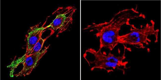
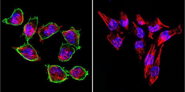
- Immunofluorescent analysis of Prolactin Receptor in H-4-II-E Cells. Cells were grown on chamber slides and fixed with formaldehyde prior to staining. Cells were probed without (control) or with a Prolactin Receptor monoclonal antibody (Product # MA1-611) at a dilution of 1:200 overnight at 4 C, washed with PBS and incubated with a DyLight-488 conjugated secondary antibody (Product # 35503). Prolactin Receptor staining (green), F-Actin staining with Phalloidin (red) and nuclei with DAPI (blue) is shown. Images were taken at 60X magnification.

- Submitted by
- Invitrogen Antibodies (provider)
- Main image

- Experimental details
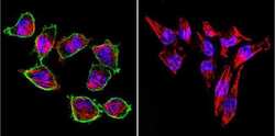
- Immunofluorescent analysis of Prolactin Receptor in SW480 Cells. Cells were grown on chamber slides and fixed with formaldehyde prior to staining. Cells were probed without (control) or with a Prolactin Receptor monoclonal antibody (Product # MA1-611) at a dilution of 1:200 overnight at 4 C, washed with PBS and incubated with a DyLight-488 conjugated secondary antibody (Product # 35503). Prolactin Receptor staining (green), F-Actin staining with Phalloidin (red) and nuclei with DAPI (blue) is shown. Images were taken at 60X magnification.

- Submitted by
- Invitrogen Antibodies (provider)
- Main image
- Experimental details
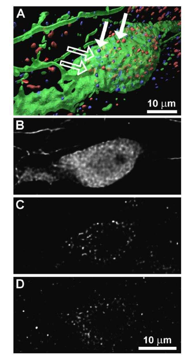
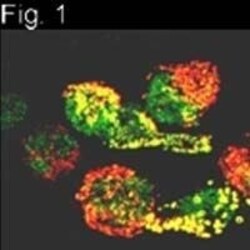
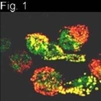
- Immunocytochemical staining of PRL receptor in NB2 cells using Product # MA1-611.

- Submitted by
- Invitrogen Antibodies (provider)
- Main image

- Experimental details
- Immunocytochemical staining of PRL receptor in NB2 cells using Product # MA1-611.
- Submitted by
- Invitrogen Antibodies (provider)
- Main image

- Experimental details
- Immunofluorescent analysis of Prolactin Receptor in C6 Cells. Cells were grown on chamber slides and fixed with formaldehyde prior to staining. Cells were probed without (control) or with a Prolactin Receptor monoclonal antibody (Product # MA1-611) at a dilution of 1:200 overnight at 4 C, washed with PBS and incubated with a DyLight-488 conjugated secondary antibody (Product # 35503). Prolactin Receptor staining (green), F-Actin staining with Phalloidin (red) and nuclei with DAPI (blue) is shown. Images were taken at 60X magnification.
- Submitted by
- Invitrogen Antibodies (provider)
- Main image

- Experimental details
- Immunofluorescent analysis of Prolactin Receptor in H-4-II-E Cells. Cells were grown on chamber slides and fixed with formaldehyde prior to staining. Cells were probed without (control) or with a Prolactin Receptor monoclonal antibody (Product # MA1-611) at a dilution of 1:200 overnight at 4 C, washed with PBS and incubated with a DyLight-488 conjugated secondary antibody (Product # 35503). Prolactin Receptor staining (green), F-Actin staining with Phalloidin (red) and nuclei with DAPI (blue) is shown. Images were taken at 60X magnification.
Supportive validation
- Submitted by
- Invitrogen Antibodies (provider)
- Main image
- Experimental details
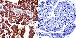
- Immunohistochemistry was performed on normal biopsies of deparaffinized Rat pituitary gland tissue. To expose target proteins, heat induced antigen retrieval was performed using 10mM sodium citrate (pH6.0) buffer, microwaved for 8-15 minutes. Following antigen retrieval tissues were blocked in 3% BSA-PBS for 30 minutes at room temperature and probed with a Prolactin Receptor monoclonal antibody (Product # MA1-611) at a dilution of 1:50 or without primary antibody (negative control) overnight at 4°C in a humidified chamber. Tissues were washed with PBST and endogenous peroxidase activity was quenched with a peroxidase suppressor. Detection was performed using a biotin-conjugated secondary antibody and SA-HRP, followed by colorimetric detection using DAB. Tissues were counterstained with hematoxylin and prepped for mounting.
- Submitted by
- Invitrogen Antibodies (provider)
- Main image

- Experimental details
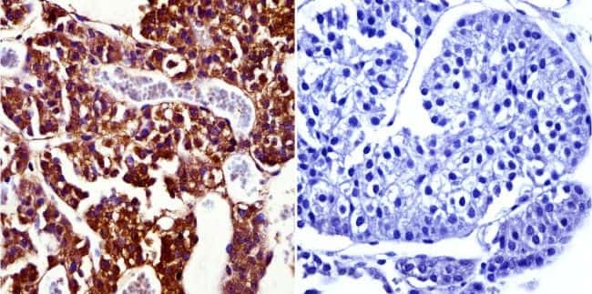
- Immunohistochemistry was performed on normal biopsies of deparaffinized Rat pituitary gland tissue. To expose target proteins, heat induced antigen retrieval was performed using 10mM sodium citrate (pH6.0) buffer, microwaved for 8-15 minutes. Following antigen retrieval tissues were blocked in 3% BSA-PBS for 30 minutes at room temperature and probed with a Prolactin Receptor monoclonal antibody (Product # MA1-611) at a dilution of 1:50 or without primary antibody (negative control) overnight at 4°C in a humidified chamber. Tissues were washed with PBST and endogenous peroxidase activity was quenched with a peroxidase suppressor. Detection was performed using a biotin-conjugated secondary antibody and SA-HRP, followed by colorimetric detection using DAB. Tissues were counterstained with hematoxylin and prepped for mounting.

Supportive validation
- Submitted by
- Invitrogen Antibodies (provider)
- Main image

- Experimental details
- NULL