 Explore
 Validate
 Learn
Learn Western blot
 Immunocytochemistry
Antibody data
- Antibody Data
- Antigen structure
- References [0]
- Comments [0]
- Validations
- Immunocytochemistry [4]
- Immunohistochemistry [1]
Submit
Validation data
Reference
Comment
Report error
- Product number
- SM5033P - Provider product page

- Provider
- Acris Antibodies GmbH
- Proper citation
- Acris Antibodies GmbH Cat#SM5033P, RRID:AB_1006631
- Product name
- anti Prolactin receptor
- Antibody type
- Monoclonal
- Antigen
- Purified Rat liver PRL receptor
- Reactivity
- Human, Rat, Porcine, Rabbit
- Host
- Mouse
- Isotype
- IgG
- Antibody clone number
- U5
- Vial size
- 0.1 mg
No comments: Submit comment
Supportive validation
- Submitted by
- Acris Antibodies GmbH (provider)
- Main image

- Experimental details
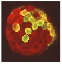
- Immunohistochemical staining of PRL receptor in rat Islets of Langerhans using SM5033P antibody.
- Submitted by
- Acris Antibodies GmbH (provider)
- Main image
- Experimental details
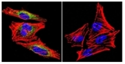
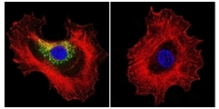
- Immunofluorescent analysis of Prolactin Receptor using Prolactin Receptor Monoclonal Antibody (U5) (Cat.-No SM5033P) shows staining in A2058 Cells. Prolactin Receptor (green), F-Actin staining with Phalloidin (red) and nuclei with DAPI (blue) is shown. Cells were grown on chamber slides and fixed with formaldehyde prior to staining. Cells were probed without (control) or with an antibody recognizing Prolactin receptor (Cat.-No SM5033P) at a dilution of 1/20 overnight at 4°C, washed with PBS and incubated with Dylight-488 conjugated secondary antibody. Images were taken 60X Magnification.

- Submitted by
- Acris Antibodies GmbH (provider)
- Main image

- Experimental details
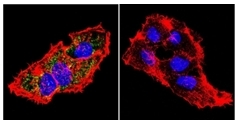
- Immunofluorescent analysis of Prolactin Receptor using Prolactin Receptor Monoclonal Antibody (U5) (Cat.-No SM5033P) shows staining in HepG2 Cells. Prolactin Receptor (green), F-Actin staining with Phalloidin (red) and nuclei with DAPI (blue) is shown. Cells were grown on chamber slides and fixed with formaldehyde prior to staining. Cells were probed without (control) or with an antibody recognizing Prolactin receptor (Cat.-No SM5033P) at a dilution of 1/20 overnight at 4°C, washed with PBS and incubated with Dylight-488 conjugated secondary antibody. Images were taken 60X Magnification.
- Submitted by
- Acris Antibodies GmbH (provider)
- Main image
- Experimental details
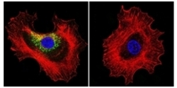
- Immunofluorescent analysis of Prolactin Receptor using Prolactin Receptor Monoclonal Antibody (U5) (Cat. SM5033P) shows staining in MCF-7 Cells. Prolactin Receptor (green), F-Actin staining with Phalloidin (red) and nuclei with DAPI (blue) is shown. Cells were grown on chamber slides and fixed with formaldehyde prior to staining. Cells were probed without (control) or with an antibody recognizing Prolactin receptor (Cat.-No SM5033P) at a dilution of 1/20 overnight at 4°C, washed with PBS and incubated with Dylight-488 conjugated secondary antibody. Images were taken 60X Magnification.
Supportive validation
- Submitted by
- Acris Antibodies GmbH (provider)
- Main image
- Experimental details
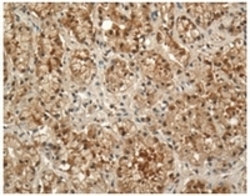
- Immunohistochemistry with anti-Prolactin Receptor Monoclonal Antibody SM5033P:Mammary gland tissue from lactating light horse mare (equine) was incubated in SM5033P at 1/100 for 1 hour at 23°C and for 24 hours at 4°C. Second antibody was Biotinylated Goat anti-Mouse IgG at 23°C for 1 hour. Development using avidin-biotin complex 23°C for 1 hour plus 3,3-diaminobenzadine for 15 min and counterstained with haematoxylin. Notes: Antigen retrieval was facilitated using steaming at 95°C for 20 min in Trizma® buffer (pH 10.0) . Blocking endogenous peroxidase performed with 0.3% H2O2 and MeOH (20 min at 23°C). To decrease non-specific staining, tissue was pre-incubated in normal goat serum for 30 min at 23°C.
