Explore
 Validate
 Learn
Learn Western blot
 Immunocytochemistry
Antibody data
- Antibody Data
- Antigen structure
- References [8]
- Comments [0]
- Validations
- Immunocytochemistry [1]
Submit
Validation data
Reference
Comment
Report error
- Product number
- HPA020923 - Provider product page

- Provider
- Atlas Antibodies
- Proper citation
- Atlas Antibodies Cat#HPA020923, RRID:AB_1852963
- Product name
- Anti-LMO7
- Antibody type
- Polyclonal
- Description
- Polyclonal Antibody against Human LMO7, Gene description: LIM domain 7, Alternative Gene Names: FBX20, FBXO20, KIAA0858, Validated applications: IHC, ICC, WB, Uniprot ID: Q8WWI1, Storage: Store at +4°C for short term storage. Long time storage is recommended at -20°C.
- Reactivity
- Human
- Host
- Rabbit
- Conjugate
- Unconjugated
- Isotype
- IgG
- Vial size
- 100 µl
- Concentration
- 0.1 mg/ml
- Storage
- Store at +4°C for short term storage. Long time storage is recommended at -20°C.
- Handling
- The antibody solution should be gently mixed before use.
Submitted references Immunoreactivity of LMO7 and other molecular markers as potential prognostic factors in oropharyngeal squamous cell carcinoma
The perinuclear region concentrates disordered proteins with predicted phase separation distributed in a 3D network of cytoskeletal filaments and organelles
New Findings on LMO7 Transcripts, Proteins and Regulatory Regions in Human and Vertebrate Model Organisms and the Intracellular Distribution in Skeletal Muscle Cells
LMO7 deficiency reveals the significance of the cuticular plate for hearing function
LRIG1‑2 and LMO7 immunoreactivity in vulvar squamous cell carcinoma: Association with prognosis in relation to HPV‑DNA and p16INK4a status
Identification of compound CA-5f as a novel late-stage autophagy inhibitor with potent anti-tumor effect against non-small cell lung cancer
The emerin-binding transcription factor Lmo7 is regulated by association with p130Cas at focal adhesions
The interactome of LIM domain proteins: The contributions of LIM domain proteins to heart failure and heart development
Israelsson P, Oda H, Öfverman C, Stefansson K, Lindquist D
BMC Oral Health 2024;24(1)
BMC Oral Health 2024;24(1)
The perinuclear region concentrates disordered proteins with predicted phase separation distributed in a 3D network of cytoskeletal filaments and organelles
do Amaral M, de Andrade Rosa I, Andrade S, Fang X, Andrade L, Costa M, Mermelstein C
Biochimica et Biophysica Acta (BBA) - Molecular Cell Research 2022;1869(1):119161
Biochimica et Biophysica Acta (BBA) - Molecular Cell Research 2022;1869(1):119161
New Findings on LMO7 Transcripts, Proteins and Regulatory Regions in Human and Vertebrate Model Organisms and the Intracellular Distribution in Skeletal Muscle Cells
Gomes G, do Amaral M, Bagri K, Vasconcellos L, Almeida M, Alvares L, Mermelstein C
International Journal of Molecular Sciences 2021;22(23):12885
International Journal of Molecular Sciences 2021;22(23):12885
LMO7 deficiency reveals the significance of the cuticular plate for hearing function
Du T, Dewey J, Wagner E, Cui R, Heo J, Park J, Francis S, Perez-Reyes E, Guillot S, Sherman N, Xu W, Oghalai J, Kachar B, Shin J
Nature Communications 2019;10(1)
Nature Communications 2019;10(1)
LRIG1‑2 and LMO7 immunoreactivity in vulvar squamous cell carcinoma: Association with prognosis in relation to HPV‑DNA and p16INK4a status
Stefansson K, Oda H, �fverman C, Lundin E, Hedman H, Lindquist D
Oncology Reports 2019
Oncology Reports 2019
Identification of compound CA-5f as a novel late-stage autophagy inhibitor with potent anti-tumor effect against non-small cell lung cancer
Zhang L, Qiang P, Yu J, Miao Y, Chen Z, Qu J, Zhao Q, Chen Z, Liu Y, Yao X, Liu B, Cui L, Jing H, Sun G
Autophagy 2018;15(3):391-406
Autophagy 2018;15(3):391-406
The emerin-binding transcription factor Lmo7 is regulated by association with p130Cas at focal adhesions
Wozniak M, Baker B, Chen C, Wilson K
PeerJ 2013;1
PeerJ 2013;1
The interactome of LIM domain proteins: The contributions of LIM domain proteins to heart failure and heart development
Li A, Ponten F, dos Remedios C
PROTEOMICS 2012;12(2):203-225
PROTEOMICS 2012;12(2):203-225
No comments: Submit comment
Supportive validation
- Submitted by
- Atlas Antibodies (provider)
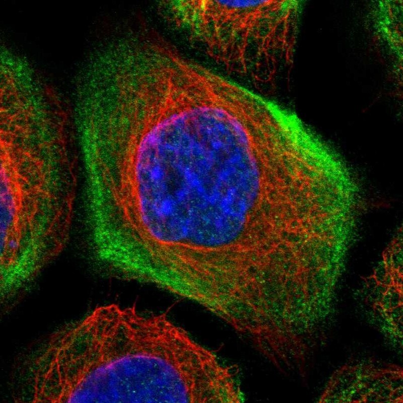
- Main image
- Experimental details
- Immunofluorescent staining of human cell line A-431 shows localization to cytosol & actin filaments.
- Sample type
- Human