Explore
 Validate
 Learn
Learn Western blot
 Immunoprecipitation
Antibody data
- Antibody Data
- Antigen structure
- References [4]
- Comments [0]
- Validations
- Immunoprecipitation [1]
- Other assay [3]
Submit
Validation data
Reference
Comment
Report error
- Product number
- PA5-72948 - Provider product page

- Provider
- Invitrogen Antibodies
- Product name
- PPARGC1A Polyclonal Antibody
- Antibody type
- Polyclonal
- Antigen
- Recombinant full-length protein
- Description
- Expected from sequence similarity: Mouse. In IHC-P, staining is very strong in the nucleus with some cytoplasmic staining. Prior to immunostaining paraffin tissues, antigen retrieval with sodium citrate buffer (pH 6.0) is recommended.
- Reactivity
- Human, Mouse, Rat, Canine, Chicken/Avian, Goat, Hamster, Porcine, Zebrafish
- Host
- Rabbit
- Isotype
- IgG
- Vial size
- 100 μL
- Concentration
- 1 mg/mL
- Storage
- Store at 4°C short term. For long term storage, store at -20°C, avoiding freeze/thaw cycles.
Submitted references Insulin-stimulated endoproteolytic TUG cleavage links energy expenditure with glucose uptake.
A novel oral nutritional supplement improves gait speed and mitochondrial functioning compared to standard care in older adults with (or at risk of) undernutrition: results from a randomized controlled trial.
Identification of transcription factor co-regulators that drive prostate cancer progression.
Whey protein sweetened with Stevia rebaudiana Bertoni (Bert.) increases mitochondrial biogenesis markers in the skeletal muscle of resistance-trained rats.
Habtemichael EN, Li DT, Camporez JP, Westergaard XO, Sales CI, Liu X, López-Giráldez F, DeVries SG, Li H, Ruiz DM, Wang KY, Sayal BS, González Zapata S, Dann P, Brown SN, Hirabara S, Vatner DF, Goedeke L, Philbrick W, Shulman GI, Bogan JS
Nature metabolism 2021 Mar;3(3):378-393
Nature metabolism 2021 Mar;3(3):378-393
A novel oral nutritional supplement improves gait speed and mitochondrial functioning compared to standard care in older adults with (or at risk of) undernutrition: results from a randomized controlled trial.
Grootswagers P, Smeets E, Oteng AB, Groot L
Aging 2021 Apr 2;13(7):9398-9418
Aging 2021 Apr 2;13(7):9398-9418
Identification of transcription factor co-regulators that drive prostate cancer progression.
Siddappa M, Wani SA, Long MD, Leach DA, Mathé EA, Bevan CL, Campbell MJ
Scientific reports 2020 Nov 23;10(1):20332
Scientific reports 2020 Nov 23;10(1):20332
Whey protein sweetened with Stevia rebaudiana Bertoni (Bert.) increases mitochondrial biogenesis markers in the skeletal muscle of resistance-trained rats.
Lima YC, Kurauti MA, da Fonseca Alves G, Ferezini J, Piovan S, Malta A, de Almeida FLA, Gomes RM, de Freitas Mathias PC, Milani PG, da Costa SC, Mareze-Costa CE
Nutrition & metabolism 2019;16:65
Nutrition & metabolism 2019;16:65
No comments: Submit comment
Supportive validation
- Submitted by
- Invitrogen Antibodies (provider)
- Main image
- Experimental details
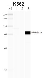
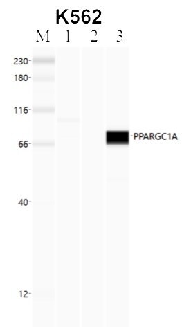
- Immunoprecipitation of PGC1 alpha was performed in K562 cells. Antigen-antibody complexes were formed by incubating approximately 500 µg whole cell lysate with 5 to 10 µL of polyclonal PGC1 alpha antibody (Product # PA5-72948) rotating 60 min at RT. The immune complexes were captured on 625 µg of anti-rabbit coated Dynabeads (Product # 11204D) and washed extensively. They were then eluted and analyzed using the Simple Western system using the same antibody as used in immunoprecipitation at a dilution of 1:25, followed by a 1:100 dilution of secondary antibody. Lane 1 is the input, lane 2 no antibody IP and lane 3 is the target specific IP. Data courtesy of the Yeo lab as part of the ENCODE project.
Supportive validation
- Submitted by
- Invitrogen Antibodies (provider)
- Main image

- Experimental details
- Immunoprecipitation of PGC1 alpha was performed in K562 cells. Antigen-antibody complexes were formed by incubating approximately 500 µg whole cell lysate with 5 to 10 µL of polyclonal PGC1 alpha antibody (Product # PA5-72948) rotating 60 min at RT. The immune complexes were captured on 625 µg of anti-rabbit coated Dynabeads (Product # 11204D) and washed extensively. They were then eluted and analyzed using the Simple Western system using the same antibody as used in immunoprecipitation at a dilution of 1:25, followed by a 1:100 dilution of secondary antibody. Lane 1 is the input, lane 2 no antibody IP and lane 3 is the target specific IP. Data courtesy of the Yeo lab as part of the ENCODE project.

- Submitted by
- Invitrogen Antibodies (provider)
- Main image

- Experimental details
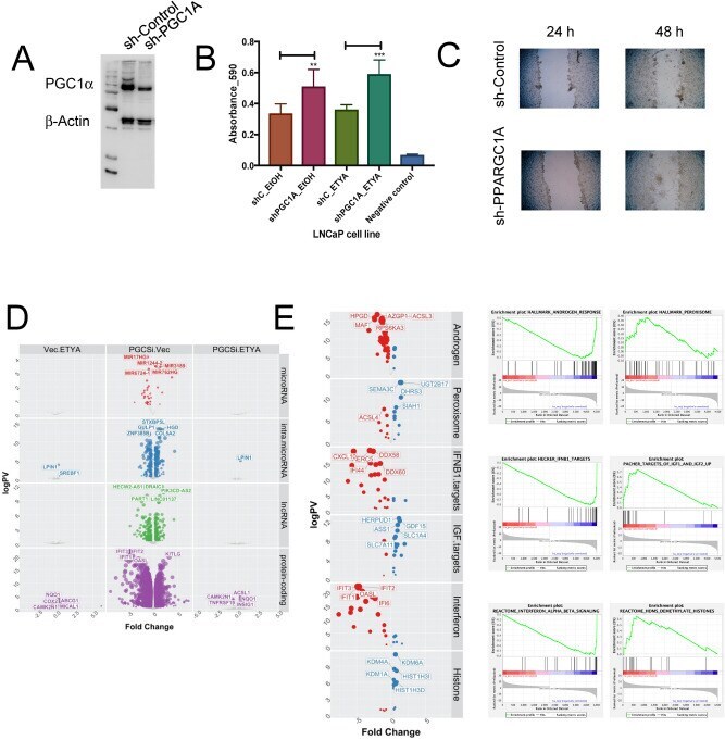
- Figure 4 Stable knockdown of PGC1alpha in LNCaP cells changes proliferation and gene expression patterns. ( A ) LNCaP cells were each stably transfected with two shRNA constructs (sh-PGC1A 34 and 35) targeting PPARGC1A resulting in reduced PGC1alpha expression (sh-PGC1A) compared to empty vector (sh-CTRL) at protein level as detected in western blotting. ( B ) Measurements of cellular levels of ATP, as an indicator of cell viability was detected in the vector controls and knockdown cells. Each measurement was performed in biological triplicates and in triplicate wells. Cells were treated in triplicate with exogenous PPARgamma ligand ETYA (10 uM, 96 hr) or EtOH vehicle control. Increased cell proliferation was seen in sh_PGC1A cells after treatment with ETYA at 96 h. ( C ) Time course scratch closure of sh-Control and sh-PPARGC1A cells mechanically wounded with p200 sterile pipette tip, sh-PPARGC1A after 48 h showed increased cell migration compared to sh-Control. ( D ) LNCaP sh-PGC1A and sh-CTRL cells were treated with ETYA (10 uM, 24 h) or EtOH vehicle control and total RNA expression and RNA-Seq undertaken according to the edgeR pipeline. Volcano plots depicting expression changes upon PGC1alpha knockdown or in response ETYA in the indicated classes of RNA in color (- log10(p.adj) > 1, abs (log2(fold change))). ( E ) Summary of significantly enriched pathways from gene set enrichment analyses (GSEA) (FDR q.val < 0.05) associated with reducing PGC1alpha expression levels.
- Submitted by
- Invitrogen Antibodies (provider)
- Main image
- Experimental details
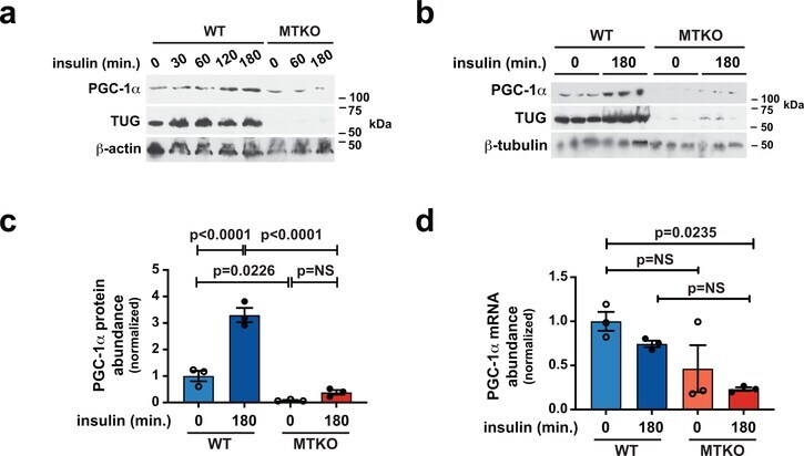
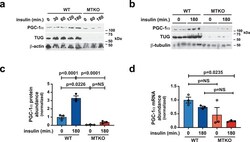
- Extended Data Fig. 7. TUG controls PGC-1alpha protein abundance. a, WT and MTKO mice were treated by IP injection of insulin-glucose, or saline control, then sacrificed at the indicated times after injection. Quadriceps muscles were immunoblotted as indicated. b,c, WT and MTKO mice were treated by IP injection of insulin-glucose, or saline control, then sacrificed after 3 h. Lysates were prepared from quadriceps muscles, PGC-1alpha was immunoblotted, and the relative abundances in each sample were quantified using densitometry. Data in (c) are presented as mean +-SEM of biologically independent samples (N=3 in each group), analyzed using ANOVA with adjustment for multiple comparisons. d, WT and MTKO mice were treated with IP insulin-glucose, or saline control, then sacrificed 3 h later. RNA was prepared from quadriceps muscles, and Q-PCR was used to measure PGC-1alpha ( Ppargc1a ) mRNA abundance. Data are presented as mean +-SEM of biologically independent samples (N=3 in each group), analyzed using ANOVA with adjustment for multiple comparisons.