Explore
 Validate
 Learn
Learn Western blot
Antibody data
- Antibody Data
- Antigen structure
- References [0]
- Comments [0]
- Validations
- Western blot [3]
- Immunocytochemistry [3]
- Immunohistochemistry [1]
Submit
Validation data
Reference
Comment
Report error
- Product number
- PA5-52602 - Provider product page

- Provider
- Invitrogen Antibodies
- Product name
- RAD18 Polyclonal Antibody
- Antibody type
- Polyclonal
- Antigen
- Recombinant full-length protein
- Description
- Immunogen sequence: LESPAKSPAS SSSKNLAVKV YTPVASRQSL KQGSRLMDNF LIREMSGSTS ELLIKENKSK FSPQKEASPA AKTKETRSVE EIAPDPSEAK RPEPPSTSTL KQVTKVDCP
- Concentration
- 0.1 mg/mL
No comments: Submit comment
Supportive validation
- Submitted by
- Invitrogen Antibodies (provider)
- Main image

- Experimental details
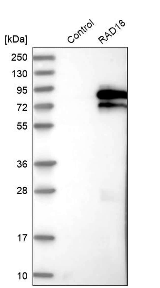
- Western blot analysis of RAD18 in control (vector only transfected HEK293T lysate) and RAD18 over-expression lysate (Co-expressed with a C-terminal myc-DDK tag (~3.1 kDa) in mammalian HEK293T cells). Samples were probed using a RAD18 Polyclonal Antibody (Product # PA5-52602).
- Submitted by
- Invitrogen Antibodies (provider)
- Main image

- Experimental details
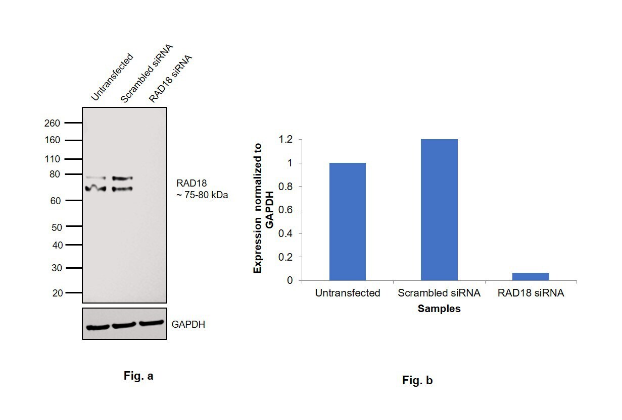
- Knockdown of RAD18 was achieved by transfecting HeLa with RAD18 specific siRNAs (Silencer® select Product # s32295 and s32296). Western blot analysis (Fig. a) was performed using Whole Cell Extract-WCL from the RAD18 knockdown cells (lane 3), non-targeting scrambled siRNA transfected cells (lane 2) and untransfected cells (lane 1). The blot was probed with RAD18 Polyclonal Antibody (Product # PA5-52602, 0.3 µg/mL) and Goat anti-Rabbit IgG (H+L) Superclonal™ Recombinant Secondary Antibody, HRP (Product # A27036, 1:4000). Densitometric analysis of this western blot is shown in histogram (Fig. b). Decrease in signal upon siRNA mediated knock down confirms that antibody is specific to RAD18.
- Submitted by
- Invitrogen Antibodies (provider)
- Main image
- Experimental details
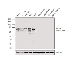
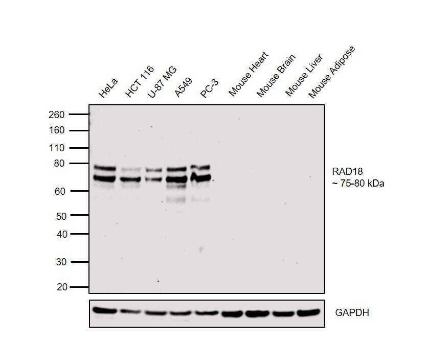
- Western blot was performed using Anti-RAD18 Polyclonal Antibody(Product # PA5-52602) and a 75 kDa and 80kDa band corresponding to RAD18 were observed across cell lines tested. This product does not show reactivity to mouse tissues. Whole Cell Extract-WCL (30 µg lysate) of HeLa (Lane 1), HCT 116 (Lane 2), U-87 MG (Lane 3), A549 (Lane 4), PC-3 (Lane 5), Mouse Heart (Lane 6), Mouse Brain (Lane 7), Mouse Liver (Lane 8) and Mouse Adipose (Lane 9) were electrophoresed using NuPAGE™ 4-12% Bis-Tris Protein Gel (Product # NP0322BOX). Resolved proteins were then transferred onto a Nitrocellulose membrane (Product # IB23001) by iBlot® 2 Dry Blotting System (Product # IB21001). The blot was probed with the primary antibody (0.4 µg/mL) and detected by chemiluminescence with Goat anti-Rabbit IgG (H+L) Superclonal™ Recombinant Secondary Antibody, HRP (Product # A27036, 1:4000) using the iBright FL 1000 (Product # A32752). Chemiluminescent detection was performed using Novex® ECL Chemiluminescent Substrate Reagent Kit (Product # WP20005).
Supportive validation
- Submitted by
- Invitrogen Antibodies (provider)
- Main image
- Experimental details
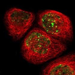
- Immunofluorescent staining of RAD18 in human cell line A-431 shows positivity in nucleus but excluded from the nucleoli. Samples were probed using a RAD18 Polyclonal Antibody (Product # PA5-52602).

- Submitted by
- Invitrogen Antibodies (provider)
- Main image

- Experimental details
- Immunofluorescent staining of RAD18 in human cell line A-431 using a RAD18 Polyclonal Antibody (Product # PA5-52602) shows localization to nucleoplasm and nuclear bodies.

- Submitted by
- Invitrogen Antibodies (provider)
- Main image

- Experimental details
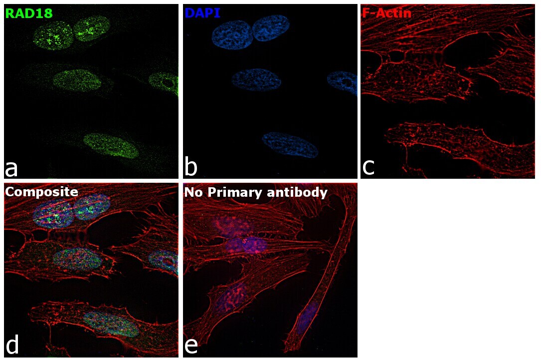
- Immunofluorescence analysis of RAD18 was performed using 70% confluent log phase HeLa cells. The cells were fixed with 4% paraformaldehyde for 10 minutes, permeabilized with 0.1% Triton™ X-100 for 15 minutes, and blocked with 2% BSA for 1 hour at room temperature. The cells were labeled with RAD18 Polyclonal Antibody (Product # PA5-52602) at 2 µg/mL in 0.1% BSA, incubated at 4 degree celsius overnight and then labeled with Goat anti-Rabbit IgG (H+L) Highly Cross-Adsorbed Secondary Antibody, Alexa Fluor Plus 488 (Product # A32731), (1:2000), for 45 minutes at room temperature (Panel a: Green). Nuclei (Panel b:Blue) were stained with ProLong™ Diamond Antifade Mountant with DAPI (Product # P36962). F-actin (Panel c: Red) was stained with Rhodamine Phalloidin (Product # R415, 1:300). Panel d represents the merged image showing nuclear localization. Panel e represents control cells with no primary antibody to assess background. The images were captured at 60X magnification.
Supportive validation
- Submitted by
- Invitrogen Antibodies (provider)
- Main image
- Experimental details
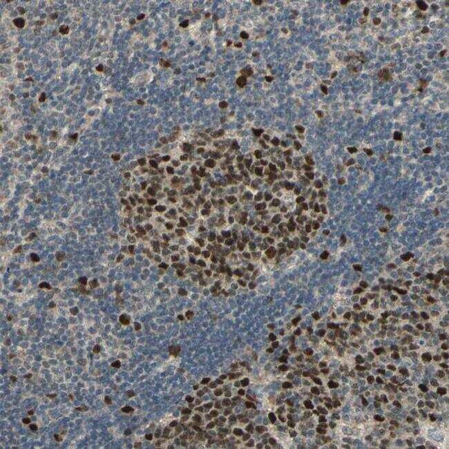
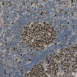
- Immunohistochemical staining of RAD18 in human lymph node shows distinct nuclear positivity in germinal center cells. Samples were probed using a RAD18 Polyclonal Antibody (Product # PA5-52602).