 Explore
 Validate
 Learn
Learn Western blot
 Immunocytochemistry
Antibody data
- Antibody Data
- Antigen structure
- References [1]
- Comments [0]
- Validations
- Immunocytochemistry [4]
- Immunohistochemistry [1]
- Flow cytometry [2]
- Other assay [1]
Submit
Validation data
Reference
Comment
Report error
- Product number
- MA5-32284 - Provider product page

- Provider
- Invitrogen Antibodies
- Product name
- RAD18 Recombinant Rabbit Monoclonal Antibody (SN66-01)
- Antibody type
- Monoclonal
- Antigen
- Synthetic peptide
- Description
- Recombinant rabbit monoclonal antibodies are produced using in vitro expression systems. The expression systems are developed by cloning in the specific antibody DNA sequences from immunoreactive rabbits. Then, individual clones are screened to select the best candidates for production. The advantages of using recombinant rabbit monoclonal antibodies include: better specificity and sensitivity, lot-to-lot consistency, animal origin-free formulations, and broader immunoreactivity to diverse targets due to larger rabbit immune repertoire.
- Reactivity
- Human, Mouse, Rat
- Host
- Rabbit
- Isotype
- IgG
- Antibody clone number
- SN66-01
- Vial size
- 100 μL
- Concentration
- 1 mg/mL
- Storage
- Store at 4°C short term. For long term storage, store at -20°C, avoiding freeze/thaw cycles.
Submitted references A positive feedback loop: RAD18-YAP-TGF-β between triple-negative breast cancer and macrophages regulates cancer stemness and progression.
Yan X, He Y, Yang S, Zeng T, Hua Y, Bao S, Yang F, Duan N, Sun C, Liang Y, Fu Z, Huang X, Li W, Yin Y
Cell death discovery 2022 Apr 12;8(1):196
Cell death discovery 2022 Apr 12;8(1):196
No comments: Submit comment
Supportive validation
- Submitted by
- Invitrogen Antibodies (provider)
- Main image
- Experimental details
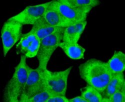
- Immunocytochemical analysis of RAD18 in Hela cells using a RAD18 Monoclonal antibody (Product # MA5-32284) as seen in green. The nuclear counter stain is DAPI (blue). Cells were fixed in paraformaldehyde, permeabilised with 0.25% Triton X100/PBS.

- Submitted by
- Invitrogen Antibodies (provider)
- Main image
- Experimental details
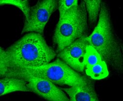
- Immunocytochemical analysis of RAD18 in NIH/3T3 cells using a RAD18 Monoclonal antibody (Product # MA5-32284) as seen in green. The nuclear counter stain is DAPI (blue). Cells were fixed in paraformaldehyde, permeabilised with 0.25% Triton X100/PBS.

- Submitted by
- Invitrogen Antibodies (provider)
- Main image
- Experimental details
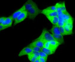
- Immunocytochemical analysis of RAD18 in 293 cells using a RAD18 Monoclonal antibody (Product # MA5-32284) as seen in green. The nuclear counter stain is DAPI (blue). Cells were fixed in paraformaldehyde, permeabilised with 0.25% Triton X100/PBS.

- Submitted by
- Invitrogen Antibodies (provider)
- Main image

- Experimental details
- Immunocytochemical analysis of RAD18 in 293 cells using a RAD18 Monoclonal antibody (Product # MA5-32284) as seen in green. The nuclear counter stain is DAPI (blue). Cells were fixed in paraformaldehyde, permeabilised with 0.25% Triton X100/PBS.

Supportive validation
- Submitted by
- Invitrogen Antibodies (provider)
- Main image
- Experimental details
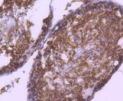
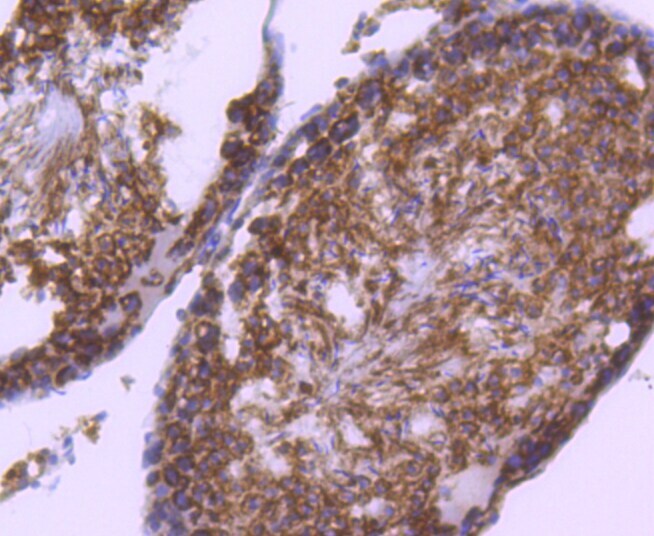
- Immunohistochemical analysis of RAD18 of paraffin-embedded rat testis tissue using a RAD18 Monoclonal antibody (Product #MA5-32284). Counter stained with hematoxylin.
Supportive validation
- Submitted by
- Invitrogen Antibodies (provider)
- Main image
- Experimental details
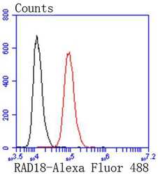
- Flow Cytometric analysis of RAD18 in Hela cells using a RAD18 Monoclonal Antibody (Product # MA5-32284) at a dilution of 1:50, as seen in red compared with an unlabelled control (cells without incubation with primary antibody; black). Alexa Fluor 488-conjugated goat anti rabbit IgG was used as the secondary antibody.

- Submitted by
- Invitrogen Antibodies (provider)
- Main image
- Experimental details
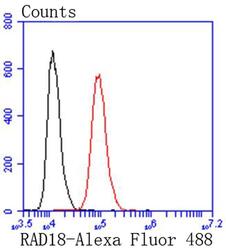
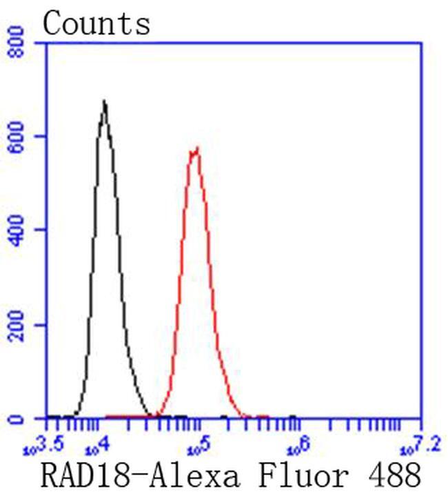
- Flow Cytometric analysis of RAD18 in Hela cells using a RAD18 Monoclonal Antibody (Product # MA5-32284) at a dilution of 1:50, as seen in red compared with an unlabelled control (cells without incubation with primary antibody; black). Alexa Fluor 488-conjugated goat anti rabbit IgG was used as the secondary antibody.
Supportive validation
- Submitted by
- Invitrogen Antibodies (provider)
- Main image
- Experimental details
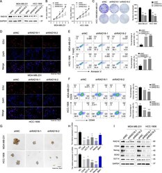
- Fig. 2 RAD18 promotes TNBC cell proliferation and maintains the stemness of TNBC in vitro. RAD18 increases TNBC cell proliferation and maintains the CSC stemness in vitro. A RAD18 protein levels in TNBC cells transfected with RAD18 shRNA lentivirus were determined by western blotting. B Cells were seeded in a 96-well plate and CCK-8 assay was used to detect cell proliferation at indicated hours (24, 48, 72, 96, and 120 h). C colony formation assay was performed to examine cell proliferation. Cells were seeded in six-well plates at a density of 2000 cells per well and cultivated for 2w. D An EdU assay was performed to determine the proliferation of different groups of TNBC cells. E Cell apoptosis in shNC and shRAD18 TNBC cells were assessed by Annexin V/7-AAD flow cytometric analysis. F Representative flow cytometric analysis of CD44+/CD24- BCSC population percentages in shNC and shRAD18 TNBC cells. G The tumor sphere formation in TNBC cells treated as indicated (200x). The quantification of mammospheres were counted using a microscope with size >=50 um. H , I mRNA and protein levels of stemness-related signature genes (CD44, OCT4, SOX2, Nanog). GAPDH was analyzed as a loading control. * p < 0.05, ** p < 0.01, *** p < 0.001. All experiments were performed independently at least three times.
