Explore
 Validate
 Learn
Learn Western blot
Antibody data
- Antibody Data
- Antigen structure
- References [0]
- Comments [0]
- Validations
- Western blot [1]
- Immunohistochemistry [13]
Submit
Validation data
Reference
Comment
Report error
- Product number
- LS-C756651 - Provider product page

- Provider
- LSBio
- Product name
- CD2AP Antibody LS-C756651
- Antibody type
- Monoclonal
- Description
- Immunogen affinity purified
- Reactivity
- Human, Mouse, Rat
- Host
- Mouse
- Isotype
- IgG
- Storage
- After reconstitution, may be stored at 4°C for 1 month. For long-term storage and to avoid freeze-thaw cycles, aliquot and store at -20°C.
No comments: Submit comment
Enhanced validation
- Submitted by
- LSBio (provider)
- Enhanced method
- Genetic validation
- Main image

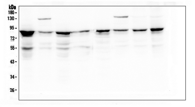
- Experimental details
- Western blot analysis of D2AP using anti-D2AP antibody. Electrophoresis was performed on a 5-20% SDS-PAGE gel at 70V (Stacking gel) / 90V (Resolving gel) for 2-3 hours. The sample well of each lane was loaded with 50ug of sample under reducing conditions. Lane 1: human K562 whole cell lysate, Lane 2: human A431 whole cell lysate, Lane 3: human 293T whole cell lysate, Lane 4: human U20S whole cell lysate, Lane 5: human HL-60 whole cell lysate, Lane 6: human MCF-7 whole cell lysate, Lane 7: human Hela whole cell lysate, Lane 8: human PANC-1 whole cell lysate. After Electrophoresis, proteins were transferred to a Nitrocellulose membrane at 150mA for 50-90 minutes. Blocked the membrane with 5% Non-fat Milk/ TBS for 1.5 hour at RT. The membrane was incubated with mouse anti-D2AP antigen affinity purified monoclonal antibody at 0.5 µg/mL overnight at 4°C, then washed with TBS-0.1% Tween 3 times with 5 minutes each and probed with a goat anti-mouse IgG-HRP secondary antibody at a dilution of 1:10000 for 1.5 hour at RT. The signal is developed using an Enhanced Chemiluminescent detection (ECL) kit with Tanon 5200 system.
Enhanced validation
- Submitted by
- LSBio (provider)
- Enhanced method
- Genetic validation
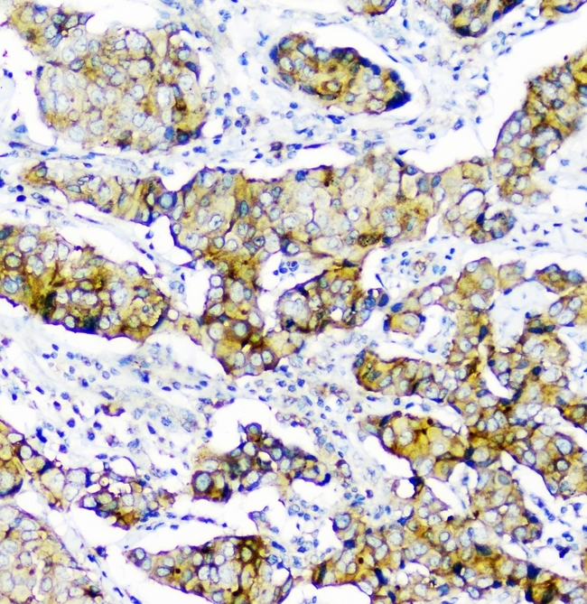
- Main image
- Experimental details
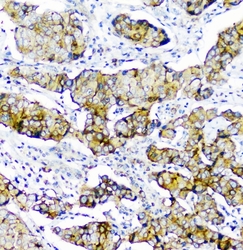
- IHC analysis of CD2AP using anti-CD2AP antibody. CD2AP was detected in paraffin-embedded section of human mammary cancer. Heat mediated antigen retrieval was performed in citrate buffer (pH6, epitope retrieval solution) for 20 mins. The tissue section was blocked with 10% goat serum. The tissue section was then incubated with 1µg/ml mouse anti-CD2AP Antibody overnight at 4°C. Biotinylated goat anti-mouse IgG was used as secondary antibody and incubated for 30 minutes at 37°C. The tissue section was developed using Strepavidin-Biotin-Complex (SABC) with DAB as the chromogen.

- Submitted by
- LSBio (provider)
- Enhanced method
- Genetic validation
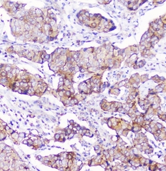
- Main image

- Experimental details
- IHC analysis of CD2AP using anti-CD2AP antibody. CD2AP was detected in paraffin-embedded section of human mammary cancer. Heat mediated antigen retrieval was performed in citrate buffer (pH6, epitope retrieval solution) for 20 mins. The tissue section was blocked with 10% goat serum. The tissue section was then incubated with 1µg/ml mouse anti-CD2AP Antibody overnight at 4°C. Biotinylated goat anti-mouse IgG was used as secondary antibody and incubated for 30 minutes at 37°C. The tissue section was developed using Strepavidin-Biotin-Complex (SABC) with DAB as the chromogen.

- Submitted by
- LSBio (provider)
- Enhanced method
- Genetic validation
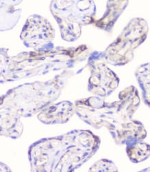
- Main image

- Experimental details
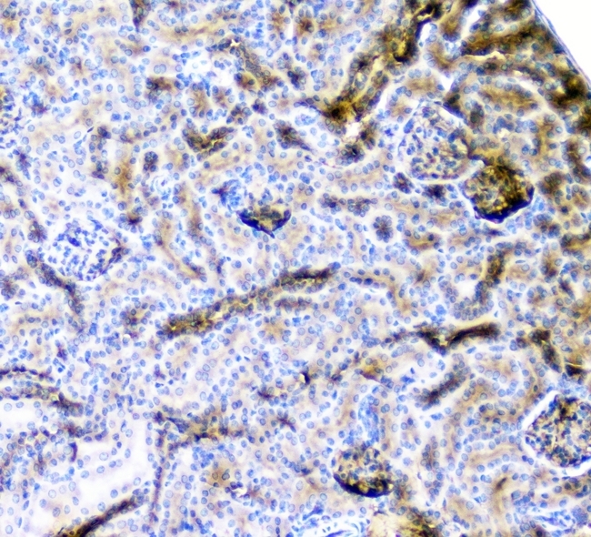
- IHC analysis of CD2AP using anti-CD2AP antibody. CD2AP was detected in paraffin-embedded section of mouse kidney tissue. Heat mediated antigen retrieval was performed in citrate buffer (pH6, epitope retrieval solution) for 20 mins. The tissue section was blocked with 10% goat serum. The tissue section was then incubated with 1µg/ml mouse anti-CD2AP Antibody overnight at 4°C. Biotinylated goat anti-mouse IgG was used as secondary antibody and incubated for 30 minutes at 37°C. The tissue section was developed using Strepavidin-Biotin-Complex (SABC) with DAB as the chromogen.

- Submitted by
- LSBio (provider)
- Enhanced method
- Genetic validation
- Main image

- Experimental details
- IHC analysis of CD2AP using anti-CD2AP antibody. CD2AP was detected in paraffin-embedded section of human mammary cancer. Heat mediated antigen retrieval was performed in citrate buffer (pH6, epitope retrieval solution) for 20 mins. The tissue section was blocked with 10% goat serum. The tissue section was then incubated with 1µg/ml mouse anti-CD2AP Antibody overnight at 4°C. Biotinylated goat anti-mouse IgG was used as secondary antibody and incubated for 30 minutes at 37°C. The tissue section was developed using Strepavidin-Biotin-Complex (SABC) with DAB as the chromogen.

- Submitted by
- LSBio (provider)
- Main image
- Experimental details
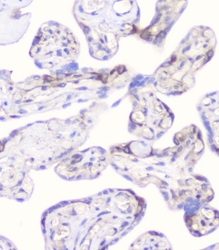
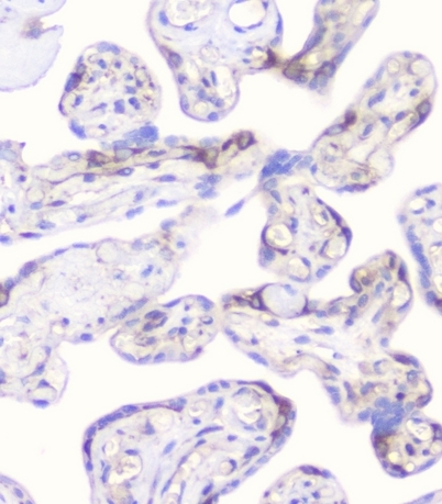
- IHC analysis of CD2AP using anti-CD2AP antibody. CD2AP was detected in paraffin-embedded section of human placenta tissue. Heat mediated antigen retrieval was performed in citrate buffer (pH6, epitope retrieval solution) for 20 mins. The tissue section was blocked with 10% goat serum. The tissue section was then incubated with 1µg/ml mouse anti-CD2AP Antibody overnight at 4°C. Biotinylated goat anti-mouse IgG was used as secondary antibody and incubated for 30 minutes at 37°C. The tissue section was developed using Strepavidin-Biotin-Complex (SABC) with DAB as the chromogen.

- Submitted by
- LSBio (provider)
- Main image

- Experimental details
- IHC analysis of CD2AP using anti-CD2AP antibody. CD2AP was detected in paraffin-embedded section of mouse kidney tissue. Heat mediated antigen retrieval was performed in citrate buffer (pH6, epitope retrieval solution) for 20 mins. The tissue section was blocked with 10% goat serum. The tissue section was then incubated with 1µg/ml mouse anti-CD2AP Antibody overnight at 4°C. Biotinylated goat anti-mouse IgG was used as secondary antibody and incubated for 30 minutes at 37°C. The tissue section was developed using Strepavidin-Biotin-Complex (SABC) with DAB as the chromogen.
- Submitted by
- LSBio (provider)
- Main image

- Experimental details
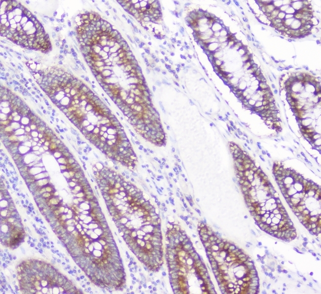
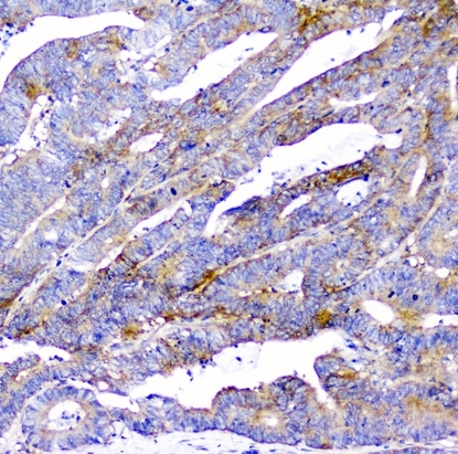
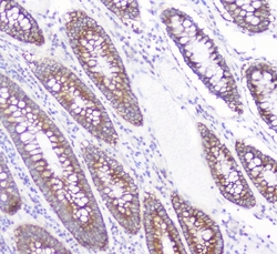
- IHC analysis of CD2AP using anti-CD2AP antibody. CD2AP was detected in paraffin-embedded section of human colon cancer. Heat mediated antigen retrieval was performed in citrate buffer (pH6, epitope retrieval solution) for 20 mins. The tissue section was blocked with 10% goat serum. The tissue section was then incubated with 1µg/ml mouse anti-CD2AP Antibody overnight at 4°C. Biotinylated goat anti-mouse IgG was used as secondary antibody and incubated for 30 minutes at 37°C. The tissue section was developed using Strepavidin-Biotin-Complex (SABC) with DAB as the chromogen.
- Submitted by
- LSBio (provider)
- Main image

- Experimental details
- IHC analysis of CD2AP using anti-CD2AP antibody. CD2AP was detected in paraffin-embedded section of human colon cancer. Heat mediated antigen retrieval was performed in citrate buffer (pH6, epitope retrieval solution) for 20 mins. The tissue section was blocked with 10% goat serum. The tissue section was then incubated with 1µg/ml mouse anti-CD2AP Antibody overnight at 4°C. Biotinylated goat anti-mouse IgG was used as secondary antibody and incubated for 30 minutes at 37°C. The tissue section was developed using Strepavidin-Biotin-Complex (SABC) with DAB as the chromogen.

- Submitted by
- LSBio (provider)
- Main image
- Experimental details
- IHC analysis of CD2AP using anti-CD2AP antibody. CD2AP was detected in paraffin-embedded section of human mammary cancer. Heat mediated antigen retrieval was performed in citrate buffer (pH6, epitope retrieval solution) for 20 mins. The tissue section was blocked with 10% goat serum. The tissue section was then incubated with 1µg/ml mouse anti-CD2AP Antibody overnight at 4°C. Biotinylated goat anti-mouse IgG was used as secondary antibody and incubated for 30 minutes at 37°C. The tissue section was developed using Strepavidin-Biotin-Complex (SABC) with DAB as the chromogen.

- Submitted by
- LSBio (provider)
- Main image
- Experimental details
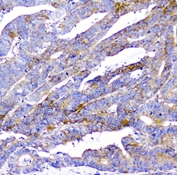
- IHC analysis of CD2AP using anti-CD2AP antibody. CD2AP was detected in paraffin-embedded section of human colon cancer. Heat mediated antigen retrieval was performed in citrate buffer (pH6, epitope retrieval solution) for 20 mins. The tissue section was blocked with 10% goat serum. The tissue section was then incubated with 1µg/ml mouse anti-CD2AP Antibody overnight at 4°C. Biotinylated goat anti-mouse IgG was used as secondary antibody and incubated for 30 minutes at 37°C. The tissue section was developed using Strepavidin-Biotin-Complex (SABC) with DAB as the chromogen.
- Submitted by
- LSBio (provider)
- Main image
- Experimental details
- IHC analysis of CD2AP using anti-CD2AP antibody. CD2AP was detected in paraffin-embedded section of human placenta tissue. Heat mediated antigen retrieval was performed in citrate buffer (pH6, epitope retrieval solution) for 20 mins. The tissue section was blocked with 10% goat serum. The tissue section was then incubated with 1µg/ml mouse anti-CD2AP Antibody overnight at 4°C. Biotinylated goat anti-mouse IgG was used as secondary antibody and incubated for 30 minutes at 37°C. The tissue section was developed using Strepavidin-Biotin-Complex (SABC) with DAB as the chromogen.
- Submitted by
- LSBio (provider)
- Main image
- Experimental details
- IHC analysis of CD2AP using anti-CD2AP antibody. CD2AP was detected in paraffin-embedded section of human colon cancer. Heat mediated antigen retrieval was performed in citrate buffer (pH6, epitope retrieval solution) for 20 mins. The tissue section was blocked with 10% goat serum. The tissue section was then incubated with 1µg/ml mouse anti-CD2AP Antibody overnight at 4°C. Biotinylated goat anti-mouse IgG was used as secondary antibody and incubated for 30 minutes at 37°C. The tissue section was developed using Strepavidin-Biotin-Complex (SABC) with DAB as the chromogen.

- Submitted by
- LSBio (provider)
- Main image

- Experimental details
- IHC analysis of CD2AP using anti-CD2AP antibody. CD2AP was detected in paraffin-embedded section of human mammary cancer. Heat mediated antigen retrieval was performed in citrate buffer (pH6, epitope retrieval solution) for 20 mins. The tissue section was blocked with 10% goat serum. The tissue section was then incubated with 1µg/ml mouse anti-CD2AP Antibody overnight at 4°C. Biotinylated goat anti-mouse IgG was used as secondary antibody and incubated for 30 minutes at 37°C. The tissue section was developed using Strepavidin-Biotin-Complex (SABC) with DAB as the chromogen.