Antibody data
- Antibody Data
- Antigen structure
- References [0]
- Comments [0]
- Validations
- Western blot [5]
- Immunocytochemistry [2]
- Immunohistochemistry [7]
- Flow cytometry [2]
Submit
Validation data
Reference
Comment
Report error
- Product number
- MA5-33009 - Provider product page
- Provider
- Invitrogen Antibodies
- Product name
- CD2AP Monoclonal Antibody (5F8)
- Antibody type
- Monoclonal
- Antigen
- Recombinant full-length protein
- Description
- Reconstitute with 0.2 mL of distilled water to yield a concentration of 500 µg/mL.
- Reactivity
- Human, Mouse, Rat
- Host
- Mouse
- Isotype
- IgG
- Antibody clone number
- 5F8
- Vial size
- 100 µg
- Concentration
- 500 µg/mL
- Storage
- Store at 4°C short term. For long term storage, store at -20°C, avoiding freeze/thaw cycles.
No comments: Submit comment
Supportive validation
- Submitted by
- Invitrogen Antibodies (provider)
- Main image
- Experimental details
- Western blot analysis of CD2AP in Lane 1: human K562 whole cell lysate, Lane 2: human A431 whole cell lysate, Lane 3: human 293T whole cell lysate, Lane 4: human U20S whole cell lysate, Lane 5: human HL-60 whole cell lysate, Lane 6: human MCF-7 whole cell lysate, Lane 7: human HeLa whole cell lysate, Lane 8: human PANC-1 whole cell lysate. Electrophoresis was performed with 5-20% SDS-PAGE gel (70V, Stacking gel; 90V Resolving gel, Time: 2-3 hours), transferred to a nitrocellulose membrane and blocked using 5% Non-fat Milk/TBS (1.5 hrs at room temperature). Samples were incubated with CD2AP monoclonal antibody (Product # MA5-33009) using a 0.5 µg/mL dilution, followed by a goat anti-mouse IgG-HRP at a dilution of 1:10,000, and developed with enhanced chemiluminescence (ECL).
- Submitted by
- Invitrogen Antibodies (provider)
- Main image
- Experimental details
- Western blot analysis of CD2AP in, Lane 1: human K562 whole cell lysate, Lane 2: human A431 whole cell lysate, Lane 3: human HEK293 whole cell lysate, Lane 4: human U20S whole cell lysate, Lane 5: human HL-60 whole cell lysate, Lane 6: human MCF-7 whole cell lysate, Lane 7: human Hela whole cell lysate, Lane 8: human PANC-1 whole cell lysate, . Electrophoresis was performed on a 5-20% SDS-PAGE gel at 70V (Stacking gel) / 90V (Resolving gel) for 2-3 hours. The sample well of each lane was loaded with 50 µg of sample under reducing conditions. After Electrophoresis, proteins were transferred to a Nitrocellulose membrane at 150mA for 50-90 minutes. The membrane was blocked with 5% Non-fat Milk/ TBS for 1. 5 hour at RT. The membrane was incubated with CD2AP Monoclonal Antibody (5F8) (Product # MA5-33009) at 0.5 μg/mL overnight at 4°C, then washed with TBS-0. 1% Tween 3 times with 5 minutes each and probed with a goat anti-mouse IgG-HRP secondary antibody at a dilution of 1:10000 for 1. 5 hour at RT. The signal is developed using an Enhanced Chemiluminescent detection (ECL) kit. A specific band was detected for CD2AP at approximately 80KD. The expected band size for CD2AP is at 71KD.
- Submitted by
- Invitrogen Antibodies (provider)
- Main image
- Experimental details
- Western blot analysis of CD2AP in, Lane 1: rat liver tissue lysate, Lane 2: rat testicular tissue lysate, Lane 3: mouse pancreas tissue lysate, Lane 4: mouse testicular tissue lysate, Lane 5: mouse RAW246. 7 whole cell lysate, . Electrophoresis was performed on a 5-20% SDS-PAGE gel at 70V (Stacking gel) / 90V (Resolving gel) for 2-3 hours. The sample well of each lane was loaded with 50 µg of sample under reducing conditions. After Electrophoresis, proteins were transferred to a Nitrocellulose membrane at 150mA for 50-90 minutes. The membrane was blocked with 5% Non-fat Milk/ TBS for 1. 5 hour at RT. The membrane was incubated with CD2AP Monoclonal Antibody (5F8) (Product # MA5-33009) at 0.5 μg/mL overnight at 4°C, then washed with TBS-0. 1% Tween 3 times with 5 minutes each and probed with a goat anti-mouse IgG-HRP secondary antibody at a dilution of 1:10000 for 1. 5 hour at RT. The signal is developed using an Enhanced Chemiluminescent detection (ECL) kit. A specific band was detected for CD2AP at approximately 80KD. The expected band size for CD2AP is at 71KD.
- Submitted by
- Invitrogen Antibodies (provider)
- Main image
- Experimental details
- Knockdown of CD2-associated protein was achieved by transfecting A-431 cells with CD2-associated protein specific siRNAs (Silencer® select Product # S24192, S24191). Western blot analysis (Fig. a) was performed using Whole cell extracts from the CD2-associated protein knockdown cells (lane 3), non-targeting scrambled siRNA transfected cells (lane 2), and untransfected cells (lane 1). The blot was probed with CD2AP Monoclonal Antibody (5F8) (Product # MA5-33009, 0.5 µg/mL) and Goat anti-Mouse IgG (H+L) Superclonal™ Recombinant Secondary Antibody, HRP (Product # A28177, 1:4000). Densitometric analysis of this western blot is shown in the histogram (Fig. b). A decrease in signal upon siRNA mediated knock down confirms that antibody is specific to CD2-associated protein.
- Submitted by
- Invitrogen Antibodies (provider)
- Main image
- Experimental details
- Western blot was performed using Anti-CD2AP Monoclonal Antibody (5F8) (Product # MA5-33009) and an 80 kDa band corresponding to CD2-associated protein was observed across cell lines tested. Whole cell extracts (30 µg lysate) of A-431 (Lane 1) and K-562 (Lane 2) were electrophoresed using NuPAGE™ 4-12% Bis-Tris Protein Gel (Product # NP0321BOX). Resolved proteins were then transferred onto a Nitrocellulose membrane (Product # IB23002) by iBlot® 2 Dry Blotting System (Product # IB21001). The blot was probed with the primary antibody (0.5 µg/mL) and detected by chemiluminescence with Goat anti-Mouse IgG (H+L) Superclonal™ Recombinant Secondary Antibody, HRP (Product # A28177, 1:4000 dilution) using the iBright FL 1000 (Product # A32752). Chemiluminescent detection was performed using SuperSignal™ West Dura Extended Duration Substrate (Product # 34076).
Supportive validation
- Submitted by
- Invitrogen Antibodies (provider)
- Main image
- Experimental details
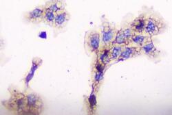
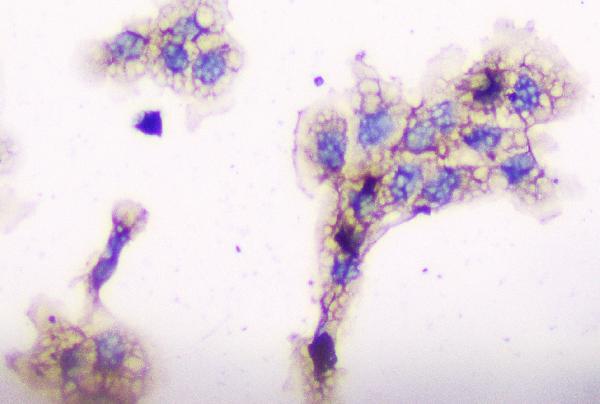
- Immunocytochemistry analysis of CD2AP using anti-CD2AP antibody (Product # MA5-33009). CD2AP was detected in a section of A431 cells. Enzyme antigen retrieval was performed using IHC enzyme antigen retrieval reagent for 15 mins. The cells were blocked with 10% goat serum and then incubated with 1μg/mL mouse anti-CD2AP antibody (Product # MA5-33009) overnight at 4°C. Biotinylated goat anti-mouse IgG was used as secondary antibody and incubated for 30 minutes at 37°C. The section was developed using Strepavidin-Biotin-Complex (SABC) with DAB as the chromogen.
- Submitted by
- Invitrogen Antibodies (provider)
- Main image
- Experimental details
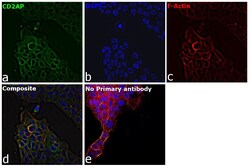
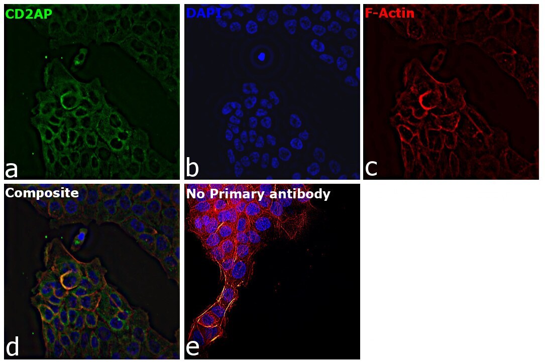
- Immunofluorescence analysis of CD2AP was performed using 70% confluent log phase A-431 cells. The cells were fixed with 4% paraformaldehyde for 10 minutes, permeabilized with 0.1% Triton™ X-100 for 15 minutes, and blocked with 2% BSA for 1 hour at room temperature. The cells were labeled with CD2AP Monoclonal Antibody (5F8) (Product # MA5-33009) at 1:100 dilution in 0.1% BSA, incubated at 4 degree celsius overnight and then with Goat anti-Mouse IgG (H+L) Highly Cross-Adsorbed Secondary Antibody, Alexa Fluor Plus 488 (Product # A32723) at a dilution of 1:2000 for 45 minutes at room temperature (Panel a: green). Nuclei (Panel b: blue) were stained with Hoechst 33342 (Product # H1399). F-actin (Panel c: red) was stained with Rhodamine Phalloidin (Product # R415, 1:300). Panel d represents the merged image showing cytoplasmic localization. Panel e represents control cells with no primary antibody to assess background. The images were captured at 40X magnification in CellInsight CX7 LZR High-Content Screening (HCS) Platform (Product # CX7C1115LZR).
Supportive validation
- Submitted by
- Invitrogen Antibodies (provider)
- Main image
- Experimental details
- Immunohistochemical analysis of CD2AP in paraffin-embedded section of human colon cancer. Heat mediated antigen retrieval was performed in citrate buffer (pH6, epitope retrieval solution) for 20 mins. The tissue section was blocked with 10% goat serum. The tissue section was then incubated with 1μg/mL mouse anti-CD2AP antibody (Product # MA5-33009) overnight at 4°C. Biotinylated goat anti-mouse IgG was used as secondary antibody and incubated for 30 minutes at 37°C. The tissue section was developed using Strepavidin-Biotin-Complex (SABC) with DAB as the chromogen.
- Submitted by
- Invitrogen Antibodies (provider)
- Main image
- Experimental details
- Immunohistochemical analysis of CD2AP in paraffin-embedded section of human mammary cancer. Heat mediated antigen retrieval was performed in citrate buffer (pH6, epitope retrieval solution) for 20 mins. The tissue section was blocked with 10% goat serum. The tissue section was then incubated with 1μg/mL mouse anti-CD2AP antibody (Product # MA5-33009) overnight at 4°C. Biotinylated goat anti-mouse IgG was used as secondary antibody and incubated for 30 minutes at 37°C. The tissue section was developed using Strepavidin-Biotin-Complex (SABC) with DAB as the chromogen.
- Submitted by
- Invitrogen Antibodies (provider)
- Main image
- Experimental details
- Immunohistochemical analysis of CD2AP in paraffin-embedded section of human placenta tissue. Heat mediated antigen retrieval was performed in citrate buffer (pH6, epitope retrieval solution) for 20 mins. The tissue section was blocked with 10% goat serum. The tissue section was then incubated with 1μg/mL mouse anti-CD2AP antibody (Product # MA5-33009) overnight at 4°C. Biotinylated goat anti-mouse IgG was used as secondary antibody and incubated for 30 minutes at 37°C. The tissue section was developed using Strepavidin-Biotin-Complex (SABC) with DAB as the chromogen.
- Submitted by
- Invitrogen Antibodies (provider)
- Main image
- Experimental details
- Immunohistochemical analysis of CD2AP in paraffin-embedded section of mouse kidney tissue. Heat mediated antigen retrieval was performed in citrate buffer (pH6, epitope retrieval solution) for 20 mins. The tissue section was blocked with 10% goat serum. The tissue section was then incubated with 1μg/mL mouse anti-CD2AP antibody (Product # MA5-33009) overnight at 4°C. Biotinylated goat anti-mouse IgG was used as secondary antibody and incubated for 30 minutes at 37°C. The tissue section was developed using Strepavidin-Biotin-Complex (SABC) with DAB as the chromogen.
- Submitted by
- Invitrogen Antibodies (provider)
- Main image
- Experimental details
- Immunohistochemical analysis of CD2AP in paraffin-embedded section of rat brain tissue. Heat mediated antigen retrieval was performed in citrate buffer (pH6, epitope retrieval solution) for 20 mins. The tissue section was blocked with 10% goat serum. The tissue section was then incubated with 1μg/mL mouse anti-CD2AP antibody (Product # MA5-33009) overnight at 4°C. Biotinylated goat anti-mouse IgG was used as secondary antibody and incubated for 30 minutes at 37°C. The tissue section was developed using Strepavidin-Biotin-Complex (SABC) with DAB as the chromogen.
- Submitted by
- Invitrogen Antibodies (provider)
- Main image
- Experimental details
- Immunohistochemical analysis of CD2AP in paraffin-embedded section of rat kidney tissue. Heat mediated antigen retrieval was performed in citrate buffer (pH6, epitope retrieval solution) for 20 mins. The tissue section was blocked with 10% goat serum. The tissue section was then incubated with 1μg/mL mouse anti-CD2AP antibody (Product # MA5-33009) overnight at 4°C. Biotinylated goat anti-mouse IgG was used as secondary antibody and incubated for 30 minutes at 37°C. The tissue section was developed using Strepavidin-Biotin-Complex (SABC) with DAB as the chromogen.
Supportive validation
- Submitted by
- Invitrogen Antibodies (provider)
- Main image
- Experimental details
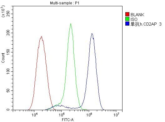
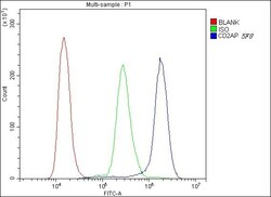
- Flow cytometry of CD2AP in K562 cells (blue line), isotype control rabbit IgG (green line) and unlabeled (red line). Samples were blocked with 10% goat serum, incubated with CD2AP monoclonal antibody (Product # MA5-33009) at a dilution of 1 µg (per 1x10^6 cells), followed by 488 conjugated goat anti-mouse IgG (30 min at 20°C) using a 5-10 µg (per 1x10^6 cells) dilution.
- Submitted by
- Invitrogen Antibodies (provider)
- Main image
- Experimental details
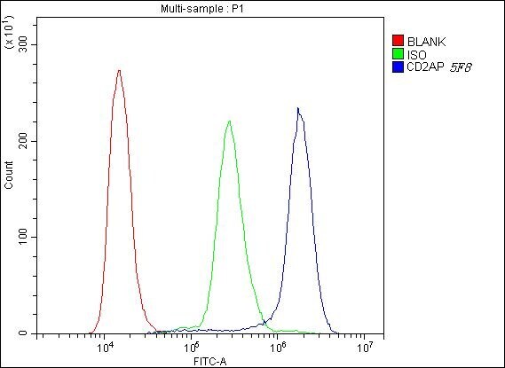
- Flow Cytometry of CD2AP in U2OS cells (blue line), isotype control mouse IgG (green line), and unlabeled (red line). Samples were blocked with 10% goat serum, incubated with CD2AP Monoclonal Antibody (5F8) (Product # MA5-33009) at a dilution of 1 μg (per 1x10^6 cells), followed by DyLight®488 conjugated goat anti-rabbit IgG (for 30 minutes at 20°C) using 5-10 μg (per 1x10^6 cells) dilution.