 Explore
 Validate
 Learn
Learn Western blot
 Immunocytochemistry
Antibody data
- Antibody Data
- Antigen structure
- References [0]
- Comments [0]
- Validations
- Western blot [3]
- Immunohistochemistry [15]
Submit
Validation data
Reference
Comment
Report error
- Product number
- NBP1-90625 - Provider product page

- Provider
- Novus Biologicals
- Proper citation
- Novus Cat#NBP1-90625, RRID:AB_11003130
- Product name
- Rabbit Polyclonal CD2AP Antibody
- Antibody type
- Polyclonal
- Description
- Immunogen affinity purified. Specificity of human CD2AP antibody verified on a Protein Array containing target protein plus 383 other non-specific proteins.
- Reactivity
- Human, Mouse, Rat
- Host
- Rabbit
- Isotype
- IgG
- Vial size
- 0.1 ml
- Storage
- Store at 4C short term. Aliquot and store at -20C long term. Avoid freeze-thaw cycles.
No comments: Submit comment
Supportive validation
- Submitted by
- Novus Biologicals (provider)
- Main image

- Experimental details
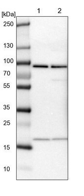
- Western Blot: CD2AP Antibody [NBP1-90625] - Lane 1: NIH-3T3 cell lysate (Mouse embryonic fibroblast cells). Lane 2: NBT-II cell lysate (Rat Wistar bladder tumor cells).
- Submitted by
- Novus Biologicals (provider)
- Main image

- Experimental details
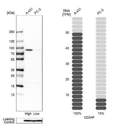
- Western Blot: CD2AP Antibody [NBP1-90625] - Analysis in human cell lines A-431 and PC-3 using anti-CD2AP antibody. Corresponding CD2AP RNA-seq data are presented for the same cell lines. Loading control: anti-PFN1.
- Submitted by
- Novus Biologicals (provider)
- Main image
- Experimental details
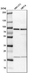
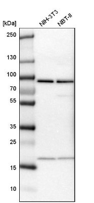
- Western Blot: CD2AP Antibody [NBP1-90625] - Analysis in mouse cell line NIH-3T3 and rat cell line NBT-II.
Supportive validation
- Submitted by
- Novus Biologicals (provider)
- Main image
- Experimental details
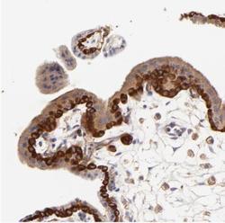
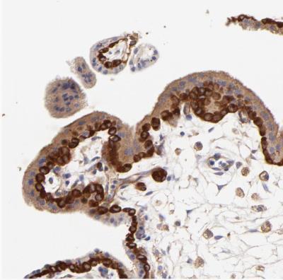
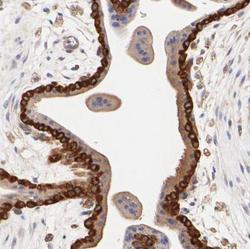
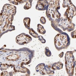
- Immunohistochemistry-Paraffin: CD2AP Antibody [NBP1-90625] - Staining of human placenta shows moderate to strong cytoplasmic positivity in trophoblastic cells.
- Submitted by
- Novus Biologicals (provider)
- Main image
- Experimental details
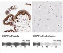
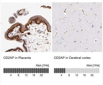
- Immunohistochemistry-Paraffin: CD2AP Antibody [NBP1-90625] - Analysis in human placenta and cerebral cortex tissues. Corresponding CD2AP RNA-seq data are presented for the same tissues.
- Submitted by
- Novus Biologicals (provider)
- Main image
- Experimental details
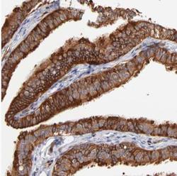
- Immunohistochemistry-Paraffin: CD2AP Antibody [NBP1-90625] - Staining of human fallopian tube shows moderate to strong cytoplasmic positivity in glandular cells.

- Submitted by
- Novus Biologicals (provider)
- Main image
- Experimental details
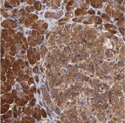
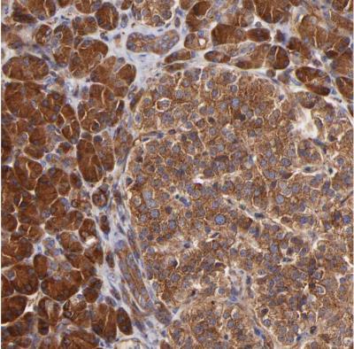
- Immunohistochemistry-Paraffin: CD2AP Antibody [NBP1-90625] - Staining of human pancreas shows moderate to strong cytoplasmic positivity in exocrine glandular cells.
- Submitted by
- Novus Biologicals (provider)
- Main image
- Experimental details
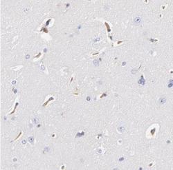
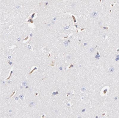
- Immunohistochemistry-Paraffin: CD2AP Antibody [NBP1-90625] - Staining of human cerebral cortex shows weak to moderate cytoplasmic positivity in endothelial cells.
- Submitted by
- Novus Biologicals (provider)
- Main image
- Experimental details
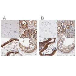
- Immunohistochemistry-Paraffin: CD2AP Antibody [NBP1-90625] - Staining of human cerebral cortex, kidney, placenta and testis using Anti-CD2AP antibody NBP1-90625 (A) shows similar protein distribution across tissues to independent antibody NBP1-90626 (B).

- Submitted by
- Novus Biologicals (provider)
- Main image

- Experimental details
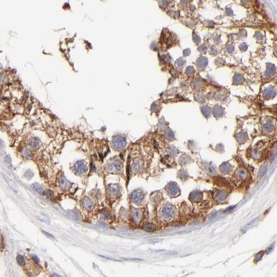
- Immunohistochemistry-Paraffin: CD2AP Antibody [NBP1-90625] - Staining of human testis.
- Submitted by
- Novus Biologicals (provider)
- Main image
- Experimental details
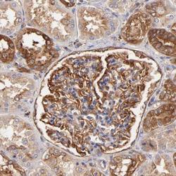
- Immunohistochemistry-Paraffin: CD2AP Antibody [NBP1-90625] - Staining of human kidney.

- Submitted by
- Novus Biologicals (provider)
- Main image
- Experimental details
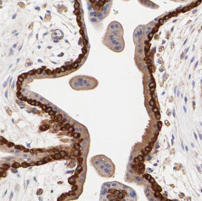
- Immunohistochemistry-Paraffin: CD2AP Antibody [NBP1-90625] - Staining of human placenta shows moderate to strong cytoplasmic positivity in trophoblastic cells.
- Submitted by
- Novus Biologicals (provider)
- Main image
- Experimental details
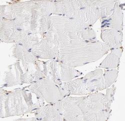
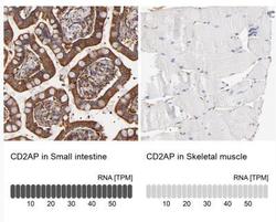
- Immunohistochemistry-Paraffin: CD2AP Antibody [NBP1-90625] - Staining of human skeletal muscle shows no positivity in myocytes.
- Submitted by
- Novus Biologicals (provider)
- Main image
- Experimental details
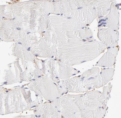
- Immunohistochemistry-Paraffin: CD2AP Antibody [NBP1-90625] - Analysis in human small intestine and skeletal muscle tissues. Corresponding CD2AP RNA-seq data are presented for the same tissues.

- Submitted by
- Novus Biologicals (provider)
- Main image
- Experimental details
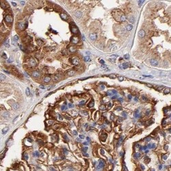
- Immunohistochemistry-Paraffin: CD2AP Antibody [NBP1-90625] - Staining of human kidney shows strong membranous positivity in cells in glomeruli.

- Submitted by
- Novus Biologicals (provider)
- Main image

- Experimental details
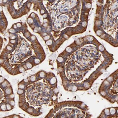
- Immunohistochemistry-Paraffin: CD2AP Antibody [NBP1-90625] - Staining of human small intestine shows strong granular positivity in cytoplasm in glandular cells.
- Submitted by
- Novus Biologicals (provider)
- Main image

- Experimental details
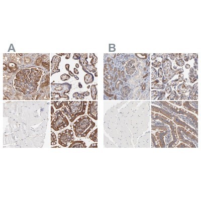
- Immunohistochemistry-Paraffin: CD2AP Antibody [NBP1-90625] - Staining of human kidney, placenta, skeletal muscle and small intestine using Anti-CD2AP antibody NBP1-90625 (A) shows similar protein distribution across tissues to independent antibody NBP1-90626 (B).
- Submitted by
- Novus Biologicals (provider)
- Main image
- Experimental details
- Immunohistochemistry-Paraffin: CD2AP Antibody [NBP1-90625] - Staining of human placenta shows strong cytoplasmic positivity in endothelial cells.
