Explore
 Validate
 Learn
Learn Western blot
 Immunocytochemistry
Antibody data
- Antibody Data
- Antigen structure
- References [6]
- Comments [0]
- Validations
- Immunocytochemistry [1]
- Immunohistochemistry [1]
Submit
Validation data
Reference
Comment
Report error
- Product number
- HPA003326 - Provider product page

- Provider
- Atlas Antibodies
- Proper citation
- Atlas Antibodies Cat#HPA003326, RRID:AB_1078457
- Product name
- Anti-CD2AP
- Antibody type
- Polyclonal
- Description
- Polyclonal Antibody against Human CD2AP, Gene description: CD2-associated protein, Alternative Gene Names: CMS, Validated applications: ICC, IHC, WB, Uniprot ID: Q9Y5K6, Storage: Store at +4°C for short term storage. Long time storage is recommended at -20°C.
- Reactivity
- Human, Mouse, Rat
- Host
- Rabbit
- Conjugate
- Unconjugated
- Isotype
- IgG
- Vial size
- 100 µl
- Concentration
- 0.2 mg/ml
- Storage
- Store at +4°C for short term storage. Long time storage is recommended at -20°C.
- Handling
- The antibody solution should be gently mixed before use.
Submitted references CD2AP deficiency aggravates Alzheimer’s disease phenotypes and pathology through p38 MAPK activation
Association of CD2AP neuronal deposits with Braak neurofibrillary stage in Alzheimer’s disease
CD2-associated protein (CD2AP) overexpression accelerates amyloid precursor protein (APP) transfer from early endosomes to the lysosomal degradation pathway
miR-29b attenuates histone deacetylase-4 mediated podocyte dysfunction and renal fibrosis in diabetic nephropathy
Podocyte injury elicits loss and recovery of cellular forces
The Alzheimer's disease risk factor CD2AP maintains blood–brain barrier integrity
Xue Y, Zhang Z, Lin R, Huang H, Zhu K, Chen D, Wu Z, Tao Q
Translational Neurodegeneration 2024;13(1)
Translational Neurodegeneration 2024;13(1)
Association of CD2AP neuronal deposits with Braak neurofibrillary stage in Alzheimer’s disease
Camacho J, Rábano A, Marazuela P, Bonaterra‐Pastra A, Serna G, Moliné T, Ramón y Cajal S, Martínez‐Sáez E, Hernández‐Guillamon M
Brain Pathology 2021;32(1)
Brain Pathology 2021;32(1)
CD2-associated protein (CD2AP) overexpression accelerates amyloid precursor protein (APP) transfer from early endosomes to the lysosomal degradation pathway
Furusawa K, Takasugi T, Chiu Y, Hori Y, Tomita T, Fukuda M, Hisanaga S
Journal of Biological Chemistry 2019;294(28):10886-10899
Journal of Biological Chemistry 2019;294(28):10886-10899
miR-29b attenuates histone deacetylase-4 mediated podocyte dysfunction and renal fibrosis in diabetic nephropathy
Gondaliya P, P. Dasare A, Jash K, Tekade R, Srivastava A, Kalia K
Journal of Diabetes & Metabolic Disorders 2019;19(1):13-27
Journal of Diabetes & Metabolic Disorders 2019;19(1):13-27
Podocyte injury elicits loss and recovery of cellular forces
Haley K, Kronenberg N, Liehm P, Elshani M, Bell C, Harrison D, Gather M, Reynolds P
Science Advances 2018;4(6)
Science Advances 2018;4(6)
The Alzheimer's disease risk factor CD2AP maintains blood–brain barrier integrity
Cochran J, Rush T, Buckingham S, Roberson E
Human Molecular Genetics 2015;24(23):6667-6674
Human Molecular Genetics 2015;24(23):6667-6674
No comments: Submit comment
Supportive validation
- Submitted by
- Atlas Antibodies (provider)
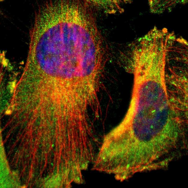
- Main image

- Experimental details
- Immunofluorescent staining of human cell line U-251 MG shows localization to plasma membrane & cytosol.
- Sample type
- Human
Supportive validation
- Submitted by
- Atlas Antibodies (provider)
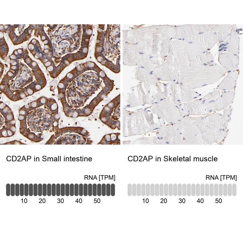
- Enhanced method
- Orthogonal validation
- Main image
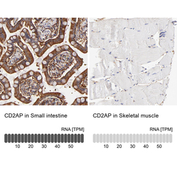
- Experimental details
- Immunohistochemistry analysis in human small intestine and skeletal muscle tissues using HPA003326 antibody. Corresponding CD2AP RNA-seq data are presented for the same tissues.
- Sample type
- Human
- Protocol
- Protocol