 Explore
 Validate
 Learn
Learn Western blot
Antibody data
- Antibody Data
- Antigen structure
- References [0]
- Comments [0]
- Validations
- Western blot [1]
- Immunohistochemistry [2]
Submit
Validation data
Reference
Comment
Report error
- Product number
- AP2163b - Provider product page

- Provider
- Abcepta
- Proper citation
- Abgent Cat#AP2163B, RRID:AB_2287705
- Product name
- TOLLIP Antibody (C-term)
- Antibody type
- Polyclonal
- Antigen
- Synthetic peptide
- Description
- Purified Rabbit Polyclonal Antibody (Pab)
- Reactivity
- Human
- Host
- Rabbit
- Isotype
- IgG
- Vial size
- 400 µl
- Concentration
- 2 mg/ml
- Storage
- Maintain refrigerated at 2-8°C for up to 6 months. For long term storage store at -20°C in small aliquots to prevent freeze-thaw cycles.
No comments: Submit comment
Supportive validation
- Submitted by
- Abcepta (provider)
- Main image
- Experimental details
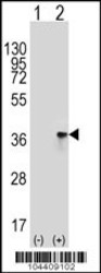
- Western blot analysis of TOLLIP (arrow) using rabbit polyclonal TOLLIP Antibody (A236) (Cat. #AP2163b). 293 cell lysates (2 ug/lane) either nontransfected (Lane 1) or transiently transfected (Lane 2) with the TOLLIP gene.
- Primary Ab dilution
- 1:1000

Supportive validation
- Submitted by
- Abcepta (provider)
- Main image

- Experimental details
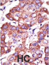
- "Formalin-fixed and paraffin-embedded human cancer tissue reacted with the primary antibody, which was peroxidase-conjugated to the secondary antibody, followed by AEC staining. This data demonstrates the use of this antibody for immunohistochemistry; clinical relevance has not been evaluated. BC = breast carcinoma; HC = hepatocarcinoma."
- Primary Ab dilution
- 1:50~100
- Submitted by
- Abcepta (provider)
- Main image
- Experimental details
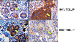
- "Middle panels, TOLLIP expression in matched normal and tumor breast samples. Here the TOLLIP stain in the invasive ductal carcinoma cells shows a much stronger signal compared with the normal ductal cells. Bottom panels, TOLLIP expression in matched normal and tumor breast samples. In this case, the signal for TOLLIP in the normal ductal cells is stronger than that in the invasive ductal carcinoma cells."
- Primary Ab dilution
- 1:50~100
