 Explore
 Validate
 Learn
Learn Western blot
 Immunocytochemistry
 Immunohistochemistry
Antibody data
- Antibody Data
- Antigen structure
- References [4]
- Comments [0]
- Validations
- Western blot [1]
- Immunocytochemistry [1]
Submit
Validation data
Reference
Comment
Report error
- Product number
- HPA038621 - Provider product page

- Provider
- Atlas Antibodies
- Proper citation
- Atlas Antibodies Cat#HPA038621, RRID:AB_10673177
- Product name
- Anti-TOLLIP
- Antibody type
- Polyclonal
- Description
- Polyclonal Antibody against Human TOLLIP, Gene description: toll interacting protein, Alternative Gene Names: IL-1RAcPIP, Validated applications: IHC, ICC, WB, Uniprot ID: Q9H0E2, Storage: Store at +4°C for short term storage. Long time storage is recommended at -20°C.
- Reactivity
- Human, Rat
- Host
- Rabbit
- Conjugate
- Unconjugated
- Isotype
- IgG
- Vial size
- 100 µl
- Concentration
- 1.0 mg/ml
- Storage
- Store at +4°C for short term storage. Long time storage is recommended at -20°C.
- Handling
- The antibody solution should be gently mixed before use.
Submitted references TOLLIP Protein Expression Predicts Unfavorable Outcome in Renal Cell Carcinoma
Toll interacting protein protects bronchial epithelial cells from bleomycin‐induced apoptosis
Endocytic Adaptor Protein Tollip Inhibits Canonical Wnt Signaling
Immunofluorescence and fluorescent-protein tagging show high correlation for protein localization in mammalian cells
Kowalewski A, Jaworski D, Borowczak J, Maniewski M, Szczerbowski K, Antosik P, Durślewicz J, Smolińska M, Ligmanowska J, Grzanka D, Szylberg Ł
International Journal of Molecular Sciences 2022;23(23):14702
International Journal of Molecular Sciences 2022;23(23):14702
Toll interacting protein protects bronchial epithelial cells from bleomycin‐induced apoptosis
Li X, Kim S, Chen T, Wang J, Yang X, Tabib T, Tan J, Guo B, Fung S, Zhao J, Sembrat J, Rojas M, Shiva S, Lafyatis R, St. Croix C, Alder J, Di Y, Kass D, Zhang Y
The FASEB Journal 2020;34(8):9884-9898
The FASEB Journal 2020;34(8):9884-9898
Endocytic Adaptor Protein Tollip Inhibits Canonical Wnt Signaling
Königshoff M, Toruń A, Szymańska E, Castanon I, Wolińska-Nizioł L, Bartosik A, Jastrzębski K, Miętkowska M, González-Gaitán M, Miaczynska M
PLOS ONE 2015;10(6):e0130818
PLOS ONE 2015;10(6):e0130818
Immunofluorescence and fluorescent-protein tagging show high correlation for protein localization in mammalian cells
Stadler C, Rexhepaj E, Singan V, Murphy R, Pepperkok R, Uhlén M, Simpson J, Lundberg E
Nature Methods 2013;10(4):315-323
Nature Methods 2013;10(4):315-323
No comments: Submit comment
Enhanced validation
- Submitted by
- Atlas Antibodies (provider)
- Enhanced method
- Genetic validation
- Main image
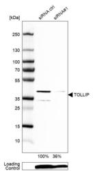
- Experimental details
- Western blot analysis in U-87MG ATCC cells transfected with control siRNA, target specific siRNA probe #1, using Anti-TOLLIP antibody. Remaining relative intensity is presented. Loading control: Anti-PPIB.
- Sample type
- Human
- Protocol
- Protocol

Supportive validation
- Submitted by
- Atlas Antibodies (provider)
- Main image
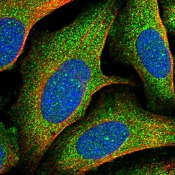
- Experimental details
- Immunofluorescent staining of human cell line U-2 OS shows localization to plasma membrane & cytosol.
- Sample type
- Human
