 Explore
 Validate
 Learn
Learn Western blot
 ELISA
ELISAAntibody data
- Antibody Data
- Antigen structure
- References [3]
- Comments [0]
- Validations
- Western blot [7]
- Immunohistochemistry [6]
Submit
Validation data
Reference
Comment
Report error
- Product number
- NBP1-77253 - Provider product page

- Provider
- Novus Biologicals
- Proper citation
- Novus Cat#NBP1-77253, RRID:AB_11009856
- Product name
- Rabbit Polyclonal XBP1 Antibody
- Antibody type
- Polyclonal
- Description
- Peptide affinity purified.
- Reactivity
- Human, Mouse, Rat
- Host
- Rabbit
- Isotype
- IgG
- Vial size
- 0.1 mg
- Concentration
- 1 mg/ml
- Storage
- Store at 4C short term. Aliquot and store at -20C long term. Avoid freeze-thaw cycles.
Submitted references MBTPS2 exacerbates albuminuria in streptozotocin-induced type I diabetic nephropathy by promoting endoplasmic reticulum stress-mediated renal damage.
Proteomics-based functional studies reveal that galectin-3 plays a protective role in the pathogenesis of intestinal Behçet's disease.
Hepatic overproduction of 13-HODE due to ALOX15 upregulation contributes to alcohol-induced liver injury in mice.
Liu Y, Xu D, Wang L, Du W, Zhang L, Xiang X
Archives of physiology and biochemistry 2020 Apr 7;:1-8
Archives of physiology and biochemistry 2020 Apr 7;:1-8
Proteomics-based functional studies reveal that galectin-3 plays a protective role in the pathogenesis of intestinal Behçet's disease.
Lee HJ, Kim JH, Hong S, Hwang I, Park SJ, Kim TI, Kim WH, Yu JW, Kim SW, Cheon JH
Scientific reports 2019 Aug 12;9(1):11716
Scientific reports 2019 Aug 12;9(1):11716
Hepatic overproduction of 13-HODE due to ALOX15 upregulation contributes to alcohol-induced liver injury in mice.
Zhang W, Zhong W, Sun Q, Sun X, Zhou Z
Scientific reports 2017 Aug 21;7(1):8976
Scientific reports 2017 Aug 21;7(1):8976
No comments: Submit comment
Supportive validation
- Submitted by
- Novus Biologicals (provider)
- Main image
- Experimental details
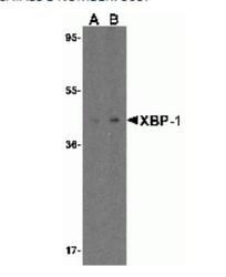
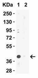
- Western Blot: XBP1 Antibody [NBP1-77253] - Analysis of XBP-1 in HepG2 cell lysate with XBP-1 antibody at 1 ug/mL in (A) the absence and (B) the presence of blocking peptide
- Submitted by
- Novus Biologicals (provider)
- Main image
- Experimental details
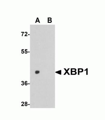
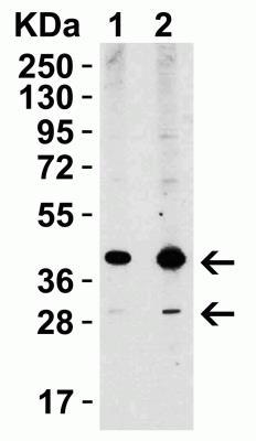
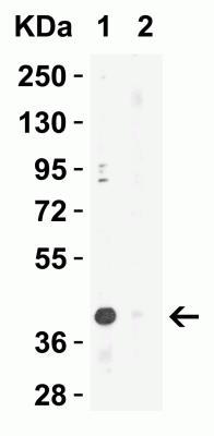
- Western Blot: XBP1 Antibody [NBP1-77253] - Analysis of XBP1 expression in rat liver tissue lysate with XBP1 antibody at (A) 1 and (B) 2 ug/ml.
- Submitted by
- Novus Biologicals (provider)
- Main image
- Experimental details
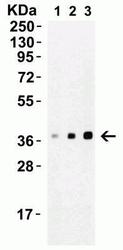
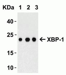
- Western Blot: XBP1 Antibody [NBP1-77253] - Loading: 15 ug of A549 cell lysate Antibodies: XBP-1, 1h incubation at RT in 5% NFDM/TBST. Secondary: Goat anti-rabbit IgG HRP conjugate at 1:10000 dilution. Lane1: 0.25 ug/mL Lane2: 0.5 ug/mL Lane3: 1 ug/mL
- Submitted by
- Novus Biologicals (provider)
- Main image

- Experimental details
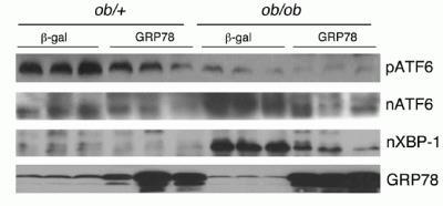
- Western Blot: XBP1 Antibody [NBP1-77253] - ob/+ and ob/ob mice were injected with an adenovirus encoding human GRP78 (Ad GRP78) and a control adenovirus encoding beta-gal (Ad beta-gal). Mature nuclear form of XBP-1 expression level was reduced in ob/ob mice with Ad GRP78 as compared to the control Ad beta-gal while there was no difference in ob/+ mice.
- Submitted by
- Novus Biologicals (provider)
- Main image
- Experimental details
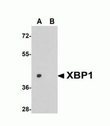
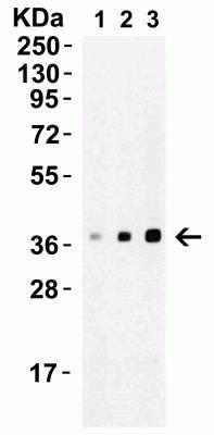
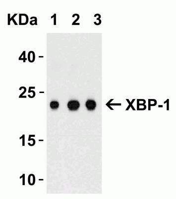
- Western Blot: XBP1 Antibody [NBP1-77253] - Loading: 15 ug of HepG2 cell lysate Antibodies: XBP-1, 1h incubation at RT in 5% NFDM/TBST. Secondary: Goat anti-rabbit IgG HRP conjugate at 1:10000 dilution. Lane1: 1 ug/mL Lane2: 2 ug/mL
- Submitted by
- Novus Biologicals (provider)
- Main image
- Experimental details
- Western Blot: XBP1 Antibody [NBP1-77253] - Loading: 100 ng of human XBP-1 recombinant protein per lane. Antibodies: XBP-1 (A: 0.5 ug/mL, B: 1 ug/mL and C: 2 ug/mL), 1h incubation at RT in 5% NFDM/TBST. Secondary: Goat anti-rabbit IgG HRP conjugate at
- Submitted by
- Novus Biologicals (provider)
- Main image
- Experimental details
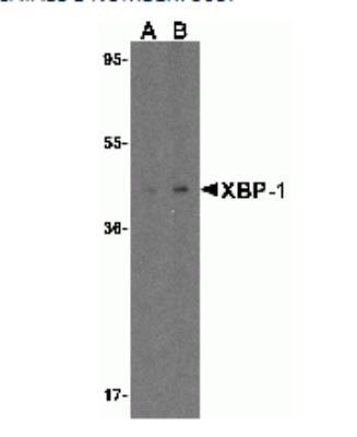
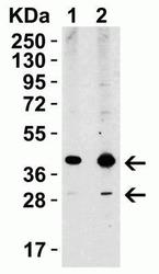
- Western Blot: XBP1 Antibody [NBP1-77253] - Loading: 15 ug of lysates per lane. Antibodies: XBP-1 (1 ug/mL), 1h incubation at RT in 5% NFDM/TBST. Secondary: Goat anti-rabbit IgG HRP conjugate at 1:10000 dilution. (1) the absence and (2) the p
Supportive validation
- Submitted by
- Novus Biologicals (provider)
- Main image
- Experimental details
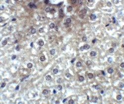
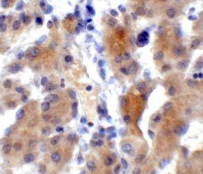
- Immunohistochemistry: XBP1 Antibody [NBP1-77253] - XBP1 in mouse liver tissue with XBP1 antibody at 2 ug/ml.
- Submitted by
- Novus Biologicals (provider)
- Main image

- Experimental details
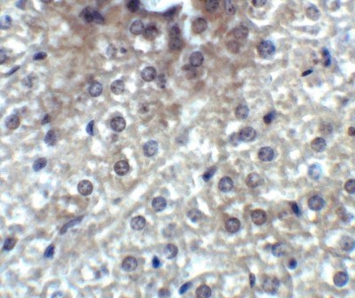
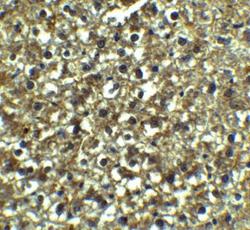
- Immunohistochemistry-Paraffin: XBP1 Antibody [NBP1-77253] - Rat liver tissue with XBP-1 Antibody at 5 ug/mL.
- Submitted by
- Novus Biologicals (provider)
- Main image

- Experimental details
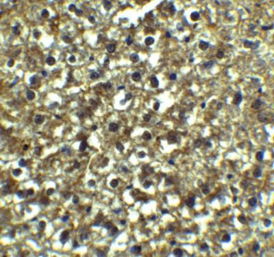
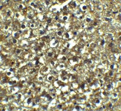
- Immunohistochemistry-Paraffin: XBP1 Antibody [NBP1-77253] - XBP1 in human liver tissue with XBP1 antibody at 5 ug/ml.
- Submitted by
- Novus Biologicals (provider)
- Main image
- Experimental details
- Immunohistochemistry-Paraffin: XBP1 Antibody [NBP1-77253] - Analysis of paraffin-embedded Rat Liver Tissue using anti-XBP-1 antibody at 5 ug/ml. Tissue was fixed with formaldehyde and blocked with 10% serum for 1 h at RT; antigen retrieval was by heat mediation with a citrate buffer (pH6). Samples were incubated with primary antibody overnight at 4C. A goat anti-rabbit IgG H&L (HRP) at 1/250 was used as secondary. Counter stained with Hematoxylin.
- Submitted by
- Novus Biologicals (provider)
- Main image
- Experimental details
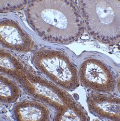
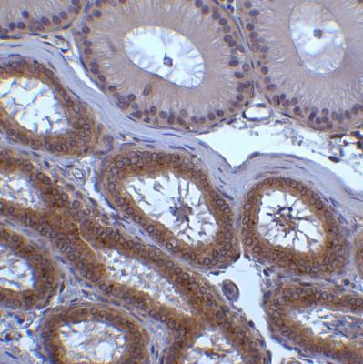
- Immunohistochemistry-Paraffin: XBP1 Antibody [NBP1-77253] - Analysis of paraffin-embedded mouse testis tissue using anti-XBP-1 antibody at 2 ug/ml. Tissue was fixed with formaldehyde and blocked with 10% serum for 1 h at RT; antigen retrieval was by heat mediation with a citrate buffer (pH6). Samples were incubated with primary antibody overnight at 4C. A goat anti-rabbit IgG H&L (HRP) at 1/250 was used as secondary. Counter stained with Hematoxylin.
- Submitted by
- Novus Biologicals (provider)
- Main image
- Experimental details
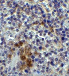
- Immunohistochemistry-Paraffin: XBP1 Antibody [NBP1-77253] - analysis of paraffin-embedded mouse spleen tissue using anti-XBP-1 antibody at 2 ug/ml. Tissue was fixed with formaldehyde and blocked with 10% serum for 1 h at RT; antigen retrieval was by heat mediation with a citrate buffer (pH6). Samples were incubated with primary antibody overnight at 4C. A goat anti-rabbit IgG H&L (HRP) at 1/250 was used as secondary. Counter stained with Hematoxylin.
