 Explore
 Validate
 Learn
Learn Western blot
Antibody data
- Antibody Data
- Antigen structure
- References [0]
- Comments [0]
- Validations
- Western blot [2]
- Immunocytochemistry [4]
- Immunohistochemistry [1]
Submit
Validation data
Reference
Comment
Report error
- Product number
- MA5-15767 - Provider product page

- Provider
- Invitrogen Antibodies
- Product name
- XBP1 Monoclonal Antibody (3H1G4)
- Antibody type
- Monoclonal
- Antigen
- Recombinant full-length protein
- Description
- A suggested positive control is XBP-1 recombinant protein.
- Reactivity
- Human, Mouse, Rat
- Host
- Mouse
- Isotype
- IgG
- Antibody clone number
- 3H1G4
- Vial size
- 100 µg
- Concentration
- 1 mg/mL
- Storage
- -20°C
No comments: Submit comment
Supportive validation
- Submitted by
- Invitrogen Antibodies (provider)
- Main image
- Experimental details
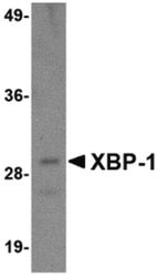
- Western blot analysis of 100 ng of XBP-1 recombinant protein using a XBP-1 monoclonal antibody (Product # MA5-15767) at 1 µg/mL.

- Submitted by
- Invitrogen Antibodies (provider)
- Main image

- Experimental details
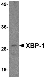
- Western Blot analysis of 100 ng of XBP-1 recombinant protein with XBP1 Monoclonal Antibody (3H1G4) (Product # MA5-15767) at 1 µg/mL.
Supportive validation
- Submitted by
- Invitrogen Antibodies (provider)
- Main image
- Experimental details
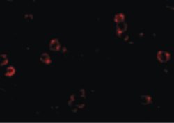
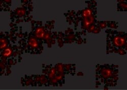
- Immunofluorescent analysis of HepG2 cells using a XBP-1 monoclonal antibody (Product # MA5-15767) at a 4 µg/mL dilution.
- Submitted by
- Invitrogen Antibodies (provider)
- Main image

- Experimental details
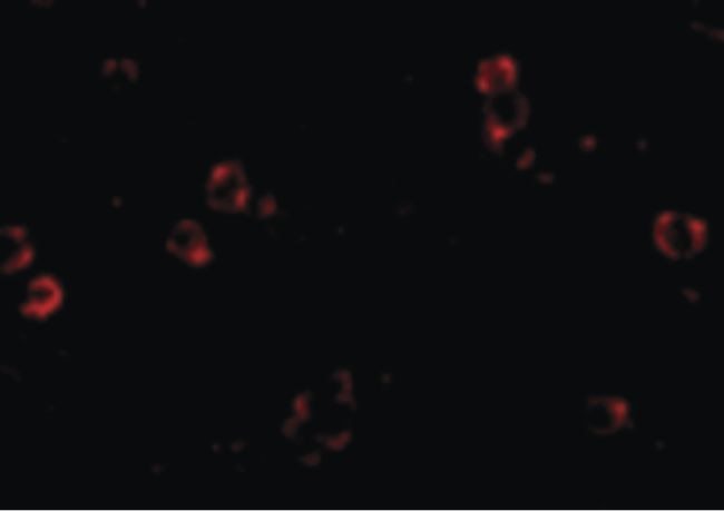
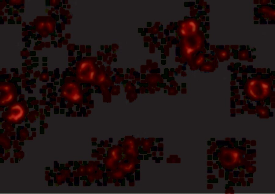
- Immunocytochemistry of XBP-1 in HepG2 cells with XBP1 Monoclonal Antibody (3H1G4) (Product # MA5-15767) at 2 µg/mL.

- Submitted by
- Invitrogen Antibodies (provider)
- Main image
- Experimental details
- Immunofluorescence of XBP1 in HepG2 cells with XBP1 Monoclonal Antibody (3H1G4) (Product # MA5-15767) at 4 µg/mL.
- Submitted by
- Invitrogen Antibodies (provider)
- Main image

- Experimental details
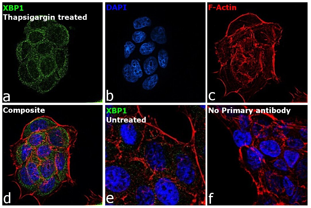
- Immunofluorescence analysis of XBP1 was performed using 70% confluent log phase MCF-7 Thapsigargin treated cells (2uM for 6hrs). The cells were fixed with 4% paraformaldehyde for 10 minutes, permeabilized with 0.1% Triton™ X-100 for 15 minutes, and blocked with 2% BSA for 45 minutes at room temperature. The cells were labeled with XBP1 Monoclonal Antibody (3H1G4) (Product # MA5-15767) at 5µg/mL in 0.1% BSA, incubated at 4 degree celsius overnight and then labeled with Donkey anti-Mouse IgG (H+L) Highly Cross-Adsorbed Secondary Antibody, Alexa Fluor Plus 488 (Product # A32766), (1:2000 dilution), for 45 minutes at room temperature (Panel a: Green). Nuclei (Panel b:Blue) were stained with ProLong™ Diamond Antifade Mountant with DAPI (Product # P36962). F-actin (Panel c: Red) was stained with Rhodamine Phalloidin (Product # R415, 1:300 dilution). Panel d represents the merged image showing cytoplasmic localization with may correspond to the unspliced variant of the protein. Panel e represents untreated cells with reduced signal. Panel f represents control cells with no primary antibody to assess background. The images were captured at 60X magnification.
Supportive validation
- Submitted by
- Invitrogen Antibodies (provider)
- Main image
- Experimental details
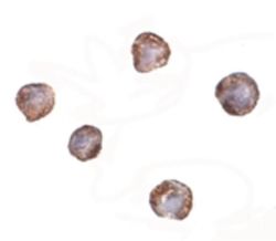
- Immunocytochemistry staining of HepG2 cells using a XBP-1 monoclonal antibody (Product # MA5-15767) at a 2 µg/mL dilution.
