Explore
 Validate
 Learn
Learn Western blot
 Immunohistochemistry
Antibody data
- Antibody Data
- Antigen structure
- References [1]
- Comments [0]
- Validations
- Immunohistochemistry [1]
- Flow cytometry [2]
- Other assay [1]
Submit
Validation data
Reference
Comment
Report error
- Product number
- PA5-25010 - Provider product page

- Provider
- Invitrogen Antibodies
- Product name
- XBP1 Polyclonal Antibody
- Antibody type
- Polyclonal
- Antigen
- Synthetic peptide
- Description
- This antibody is predicted to react with bovine, mouse and rat based on sequence homology.
- Reactivity
- Human
- Host
- Rabbit
- Isotype
- IgG
- Vial size
- 400 μL
- Concentration
- 0.5 mg/mL
- Storage
- Store at 4°C short term. For long term storage, store at -20°C, avoiding freeze/thaw cycles.
Submitted references Detection of endoplasmic reticulum stress and the unfolded protein response in naturally-occurring endocrinopathic equine laminitis.
Cassimeris L, Engiles JB, Galantino-Homer H
BMC veterinary research 2019 Jan 10;15(1):24
BMC veterinary research 2019 Jan 10;15(1):24
No comments: Submit comment
Supportive validation
- Submitted by
- Invitrogen Antibodies (provider)
- Main image
- Experimental details
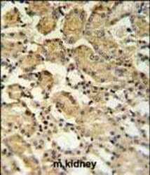
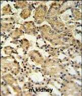
- Immunohistochemistry analysis of XBP1 in formalin fixed and paraffin embedded mouse kidney tissue. Samples were incubated with XBP1 polyclonal antibody (Product # PA5-25010) followed by peroxidase conjugation of the secondary antibody and DAB staining. This data demonstrates the use of this antibody for immunohistochemistry. Clinical relevance has not been evaluated.
Supportive validation
- Submitted by
- Invitrogen Antibodies (provider)
- Main image
- Experimental details
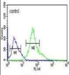
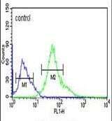
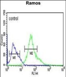
- Flow cytometry analysis of Ramos cells using a XBP1 polyclonal antibody (Product # PA5-25010) (right) compared to a negative control cell (left) at a dilution of 1:10-50, followed by a FITC-conjugated goat anti-rabbit antibody
- Submitted by
- Invitrogen Antibodies (provider)
- Main image
- Experimental details
- Flow cytometry of XBP1 in Ramos cells (right histogram). Samples were incubated with XBP1 polyclonal antibody (Product # PA5-25010) followed by FITC-conjugated goat-anti-rabbit secondary antibody. Negative control cell (left histogram).

Supportive validation
- Submitted by
- Invitrogen Antibodies (provider)
- Main image
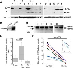
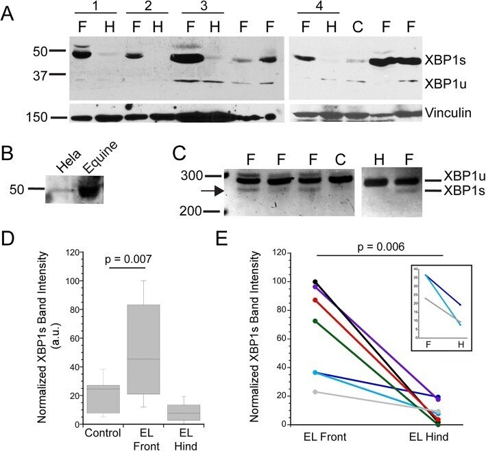
- Experimental details
- Fig. 1 Spliced XBP1 is expressed in laminitic tissues. a . Representative immunoblots probed for expression of the XBP1s or XBP1u proteins derived from spliced and unspliced XBP1 mRNA, respectively. XBP1s is prevalent in EL front limbs (F) and barely detectable in either EL hind limbs (H) or controls (C). Membranes were reprobed for vinculin as a loading control. Four pairs of EL front and hind limbs from the same horses are noted by horizontal bars and numbered 1-4 (pair 1: 116 LF, RH; pair 2: 141 LF, RH; pair 3: 134 RF, RH; pair 4: 104 RF, 104 RH; see Table 1 ). b . The antibody to XBP1 recognized a band of the same mw in both Hela (human) cells and equine lamellar tissue. c PCR amplification of a region near the XBP1 splice site produces amplimers of the expected base pairs for spliced and unspliced XBP1. Samples are labeled as in ( a ). d , e . Immunoblot band intensities for XBP1s were measured and normalized as described in Methods. d Box blot summarizing XBP1s expression levels in control ( n = 7), EL front ( n = 12) and EL hind limbs ( n = 7). Horizontal lines represent the median value for each group. XBP1s expression was not significantly different between control and EL hind limbs ( p = 0.08). e Comparison of XBP1s expression levels in paired samples from EL front and EL hind limbs of the same horses ( n = 7 pairs), inset shows the 3 sample pairs with similar XBP1s band intensities between front (F) and hind limb (H). Although the differences between front and hind