Explore
 Validate
 Learn
Learn Western blot
 Immunocytochemistry
Antibody data
- Antibody Data
- Antigen structure
- References [3]
- Comments [0]
- Validations
- Immunocytochemistry [4]
- Immunohistochemistry [2]
- Other assay [3]
Submit
Validation data
Reference
Comment
Report error
- Product number
- PA5-11274 - Provider product page

- Provider
- Invitrogen Antibodies
- Product name
- IFITM3 Polyclonal Antibody
- Antibody type
- Polyclonal
- Antigen
- Synthetic peptide
- Reactivity
- Human, Mouse
- Host
- Rabbit
- Isotype
- IgG
- Vial size
- 400 μL
- Concentration
- 0.47 mg/mL
- Storage
- Store at 4°C short term. For long term storage, store at -20°C, avoiding freeze/thaw cycles.
Submitted references Increased Expression of Interferon-Induced Transmembrane 3 (IFITM3) in Stroke and Other Inflammatory Conditions in the Brain.
Cutting Edge: NOX2 NADPH Oxidase Controls Infection by an Intracellular Bacterial Pathogen through Limiting the Type 1 IFN Response.
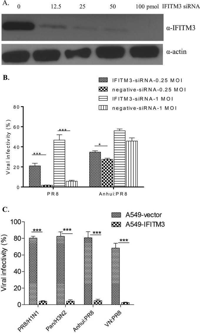
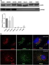
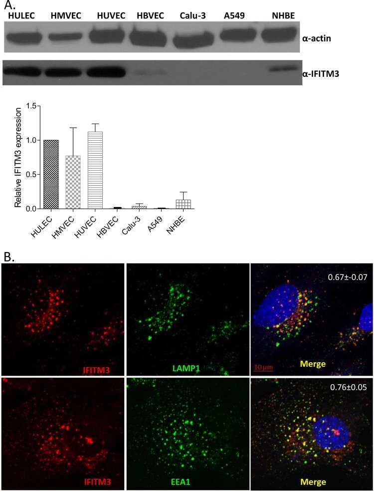
Constitutively Expressed IFITM3 Protein in Human Endothelial Cells Poses an Early Infection Block to Human Influenza Viruses.
Harmon E, Doan A, Bautista-Garrido J, Jung JE, Marrelli SP, Kim GS
International journal of molecular sciences 2022 Aug 10;23(16)
International journal of molecular sciences 2022 Aug 10;23(16)
Cutting Edge: NOX2 NADPH Oxidase Controls Infection by an Intracellular Bacterial Pathogen through Limiting the Type 1 IFN Response.
Rojas Márquez JD, Li T, McCluggage ARR, Tan JMJ, Muise A, Higgins DE, Brumell JH
Journal of immunology (Baltimore, Md. : 1950) 2021 Jan 15;206(2):323-328
Journal of immunology (Baltimore, Md. : 1950) 2021 Jan 15;206(2):323-328
Constitutively Expressed IFITM3 Protein in Human Endothelial Cells Poses an Early Infection Block to Human Influenza Viruses.
Sun X, Zeng H, Kumar A, Belser JA, Maines TR, Tumpey TM
Journal of virology 2016 Dec 15;90(24):11157-11167
Journal of virology 2016 Dec 15;90(24):11157-11167
No comments: Submit comment
Supportive validation
- Submitted by
- Invitrogen Antibodies (provider)
- Main image

- Experimental details
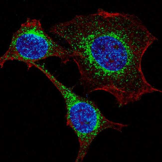
- Immunofluorescent analysis of HeLa cells stained with an IFITM3 polyclonal antibody (Product # PA5-11274). HeLa cells were fixed with 4% PFA (20 min), permeabilized with Triton X-100 (0.2%, 30 min), then incubated with an IFITM3 polyclonal antibody (Product # PA5-11274) (1:200, 2 hr at room temperature). Primary antibody detected by fluor-conjugated donkey anti-rabbit secondary antibody (green) was used (1:1000, 1hr). Cytoplasmic actin was counterstained with red dye conjugated phalloidin (5.25 uM, 25 min). Nuclei were counterstained with Hoechst 33342 (blue) (10 µg/mL, 3 min)
- Submitted by
- Invitrogen Antibodies (provider)
- Main image
- Experimental details
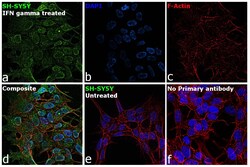
- Immunofluorescence analysis of Interferon-induced transmembrane protein 3 was performed using 70% confluent log phase SH-SY5Y IFN gamma, 100 units/mL for 24 hrs. The cells were fixed with 4% paraformaldehyde for 10 minutes, permeabilized with 0.1% Triton™ X-100 for 15 minutes, and blocked with 2% BSA for 1 hour at room temperature. The cells were labeled with IFITM3 Polyclonal Antibody (Product # PA5-11274) at 1:100 in 0.1% BSA, incubated at 4 degree celsius overnight and then labeled with Donkey anti-Rabbit IgG (H+L) Highly Cross-Adsorbed Secondary Antibody, Alexa Fluor Plus 488 (Product # A32790), (1:2000), for 45 minutes at room temperature (Panel a: Green). Nuclei (Panel b:Blue) were stained with ProLong™ Diamond Antifade Mountant with DAPI (Product # P36962). F-actin (Panel c: Red) was stained with Rhodamine Phalloidin (Product # R415, 1:300). Panel d represents the merged image showing Cytoplasmic localization. Panel e represents no signal was observed Panel f represents control cells with no primary antibody to assess background. The images were captured at 60X magnification.

- Submitted by
- Invitrogen Antibodies (provider)
- Main image

- Experimental details
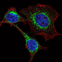
- Immunofluorescence analysis of Interferon-induced transmembrane protein 3 was performed using 70% confluent log phase SH-SY5Y IFN gamma, 100 units/mL for 24 hrs. The cells were fixed with 4% paraformaldehyde for 10 minutes, permeabilized with 0.1% Triton™ X-100 for 15 minutes, and blocked with 2% BSA for 1 hour at room temperature. The cells were labeled with IFITM3 Polyclonal Antibody (Product # PA5-11274) at 1:100 in 0.1% BSA, incubated at 4 degree celsius overnight and then labeled with Donkey anti-Rabbit IgG (H+L) Highly Cross-Adsorbed Secondary Antibody, Alexa Fluor Plus 488 (Product # A32790), (1:2000), for 45 minutes at room temperature (Panel a: Green). Nuclei (Panel b:Blue) were stained with ProLong™ Diamond Antifade Mountant with DAPI (Product # P36962). F-actin (Panel c: Red) was stained with Rhodamine Phalloidin (Product # R415, 1:300). Panel d represents the merged image showing Cytoplasmic localization. Panel e represents no signal was observed Panel f represents control cells with no primary antibody to assess background. The images were captured at 60X magnification.

- Submitted by
- Invitrogen Antibodies (provider)
- Main image
- Experimental details
- Immunocytochemistry analysis of IFITM3 in HeLa cells. Samples were incubated with IFITM3 polyclonal antibody (Product # PA5-11274) using a dilution of 1:200 for 2 h at room temperature followed by Alexa Fluor® 488 conjugated donkey anti-rabbit at a dilution of 1:1,000 for 1hr. Cells were fixed with 4% PFA (20 min) and permeabilized with Triton X-100 (0.2%, 30 min). Cytoplasmic actin was counterstained with Alexa Fluor® 555 (red) conjugated Phalloidin (5.25 μM, 25 min). Nuclei were counterstained with Hoechst 33342 (blue) (10 µg/mL, 3 min). Note the highly specific localization of the IFITM3 immunoreactivity to the Golgi.

Supportive validation
- Submitted by
- Invitrogen Antibodies (provider)
- Main image
- Experimental details
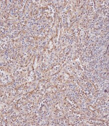
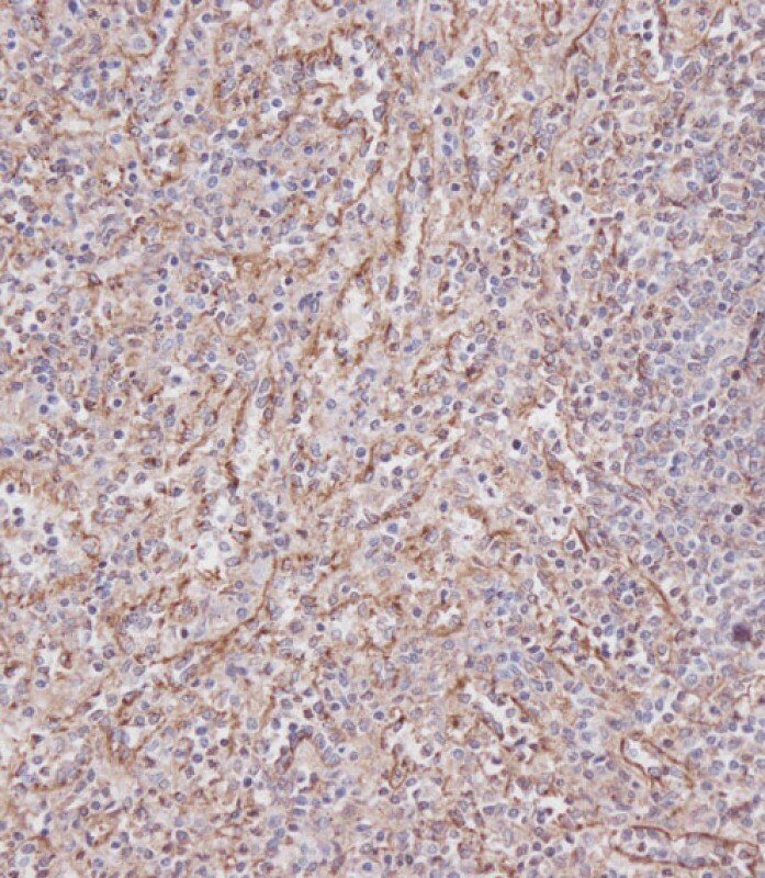
- Immunohistochemistry analysis of IFITM3 in paraffin-embedded Human spleen tissue. Samples were incubated with IFITM3 polyclonal antibody (Product # PA5-11274) using a dilution of 1:100 for 15 min at room temperature followed by Leica Bond Polymer Refine Detection. Tissue was fixed with formaldehyde at room temperature. Heat induced epitope retrieval was performed by EDTA buffer (pH 9 0).
- Submitted by
- Invitrogen Antibodies (provider)
- Main image
- Experimental details
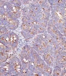
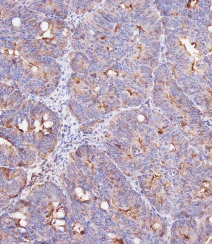
- Immunohistochemistry analysis of IFITM3 in paraffin-embedded Human colon carcinoma tissue. Samples were incubated with IFITM3 polyclonal antibody (Product # PA5-11274) using a dilution of 1:100 for 15 min at room temperature followed by Leica Bond Polymer Refine Detection. Tissue was fixed with formaldehyde at room temperature. Heat induced epitope retrieval was performed by EDTA buffer (pH 9 0).
Supportive validation
- Submitted by
- Invitrogen Antibodies (provider)
- Main image

- Experimental details
- NULL
- Submitted by
- Invitrogen Antibodies (provider)
- Main image
- Experimental details
- NULL
- Submitted by
- Invitrogen Antibodies (provider)
- Main image

- Experimental details
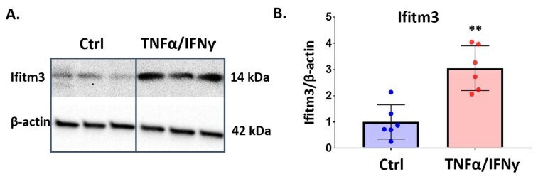
- The IFITM3 protein level is increased by TNFalpha and IFNgamma in primary microglia. Primary microglia were treated with TNFalpha and IFNgamma (20 ng/mL) for 24 h. Western blotting was performed to measure protein level of IFITM3. ( A ) IFITM3 (seen at 14 kDa) was increased in the treatment group, compared to control. ( B ) Quantification of IFITM3 to beta-actin showed the significant induction of IFITM3 protein in microglia ( n = 6, ** p < 0.01, two-tailed unpaired Student's t -test).