 Explore
 Validate
 Learn
Learn Western blot
Antibody data
- Antibody Data
- Antigen structure
- References [2]
- Comments [0]
- Validations
- Western blot [3]
- Immunocytochemistry [1]
- Immunohistochemistry [1]
Submit
Validation data
Reference
Comment
Report error
- Product number
- GTX118732 - Provider product page

- Provider
- GeneTex
- Proper citation
- GeneTex Cat#GTX118732, RRID:AB_10618597
- Product name
- HAUS6 antibody
- Antibody type
- Polyclonal
- Reactivity
- Human
- Host
- Rabbit
Submitted references Mitotic spindle assembly and γ-tubulin localisation depend on the integral nuclear membrane protein Samp1.
Aurora-A phosphorylates Augmin complex component Hice1 protein at an N-terminal serine/threonine cluster to modulate its microtubule binding activity during spindle assembly.
Larsson VJ, Jafferali MH, Vijayaraghavan B, Figueroa RA, Hallberg E
Journal of cell science 2018 Apr 13;131(8)
Journal of cell science 2018 Apr 13;131(8)
Aurora-A phosphorylates Augmin complex component Hice1 protein at an N-terminal serine/threonine cluster to modulate its microtubule binding activity during spindle assembly.
Tsai CY, Ngo B, Tapadia A, Hsu PH, Wu G, Lee WH
The Journal of biological chemistry 2011 Aug 26;286(34):30097-106
The Journal of biological chemistry 2011 Aug 26;286(34):30097-106
No comments: Submit comment
Supportive validation
- Submitted by
- GeneTex (provider)
- Main image

- Experimental details
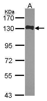
- Sample (30 ?g of whole cell lysate) A: JurKat 7.5% SDS PAGE GTX118732 diluted at 1:1000 The HRP-conjugated anti-rabbit IgG antibody (GTX213110-01) was used to detect the primary antibody.
- Submitted by
- GeneTex (provider)
- Main image
- Experimental details
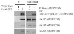
- IP-WB assay to show that Hice1 (GTX113617) co-immunoprecipitated with other Augmin components HAUS6 (GTX118732), HAUS2 (GTX118734) and HAUS1 (GTX118733) in U2OS cells. The HRP-conjugated anti-rabbit IgG antibody (GTX213110-01) was used to detect the primary antibody.

- Submitted by
- GeneTex (provider)
- Main image
- Experimental details
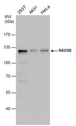
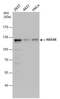
- HAUS6 antibody detects HAUS6 protein by western blot analysis. Various whole cell extracts (30 ?g) were separated by 7.5% SDS-PAGE, and the membrane was blotted with HAUS6 antibody (GTX118732) diluted by 1:1000. The HRP-conjugated anti-rabbit IgG antibody (GTX213110-01) was used to detect the primary antibody.
Supportive validation
- Submitted by
- GeneTex (provider)
- Main image
- Experimental details
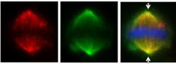
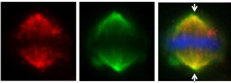
- HAUS6 ( IFA using GTX118732 at 1: 500; red) co-localizes at the mitotic spindle with Hice1-GFP (HAUS-GFP; green) in U2OS cells. DAPI staining (blue) shows mitotic chromosomes. Arrows indicate spindle poles.
Supportive validation
- Submitted by
- GeneTex (provider)
- Main image
- Experimental details
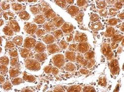
- HAUS6 antibody detects HAUS6 protein on human colon carcinoma by immunohistochemical analysis. Sample: Paraffin-embedded colon carcinoma. HAUS6 antibody (GTX118732) dilution: 1:500.
