Explore
 Validate
 Learn
Learn Immunocytochemistry
Antibody data
- Antibody Data
- Antigen structure
- References [3]
- Comments [0]
- Validations
- Immunocytochemistry [1]
- Immunohistochemistry [1]
Submit
Validation data
Reference
Comment
Report error
- Product number
- HPA024446 - Provider product page

- Provider
- Atlas Antibodies
- Proper citation
- Atlas Antibodies Cat#HPA024446, RRID:AB_1848680
- Product name
- Anti-VPS53
- Antibody type
- Polyclonal
- Description
- Polyclonal Antibody against Human VPS53, Gene description: vacuolar protein sorting 53 homolog (S. cerevisiae), Alternative Gene Names: FLJ10979, HCCS1, Validated applications: ICC, IHC, Uniprot ID: Q5VIR6, Storage: Store at +4°C for short term storage. Long time storage is recommended at -20°C.
- Reactivity
- Human
- Host
- Rabbit
- Conjugate
- Unconjugated
- Isotype
- IgG
- Vial size
- 100 µl
- Concentration
- 0.2 mg/ml
- Storage
- Store at +4°C for short term storage. Long time storage is recommended at -20°C.
- Handling
- The antibody solution should be gently mixed before use.
Submitted references ARFRP1 functions upstream of ARL1 and ARL5 to coordinate recruitment of distinct tethering factors to the trans-Golgi network.
A neurodevelopmental disorder caused by mutations in the VPS51 subunit of the GARP and EARP complexes.
Proteomic and Biochemical Comparison of the Cellular Interaction Partners of Human VPS33A and VPS33B
Ishida M, Bonifacino JS
The Journal of cell biology 2019 Nov 4;218(11):3681-3696
The Journal of cell biology 2019 Nov 4;218(11):3681-3696
A neurodevelopmental disorder caused by mutations in the VPS51 subunit of the GARP and EARP complexes.
Gershlick DC, Ishida M, Jones JR, Bellomo A, Bonifacino JS, Everman DB
Human molecular genetics 2019 May 1;28(9):1548-1560
Human molecular genetics 2019 May 1;28(9):1548-1560
Proteomic and Biochemical Comparison of the Cellular Interaction Partners of Human VPS33A and VPS33B
Hunter M, Hesketh G, Benedyk T, Gingras A, Graham S
Journal of Molecular Biology 2018;430(14):2153-2163
Journal of Molecular Biology 2018;430(14):2153-2163
No comments: Submit comment
Supportive validation
- Submitted by
- Atlas Antibodies (provider)
- Main image
- Experimental details
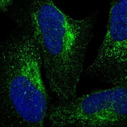
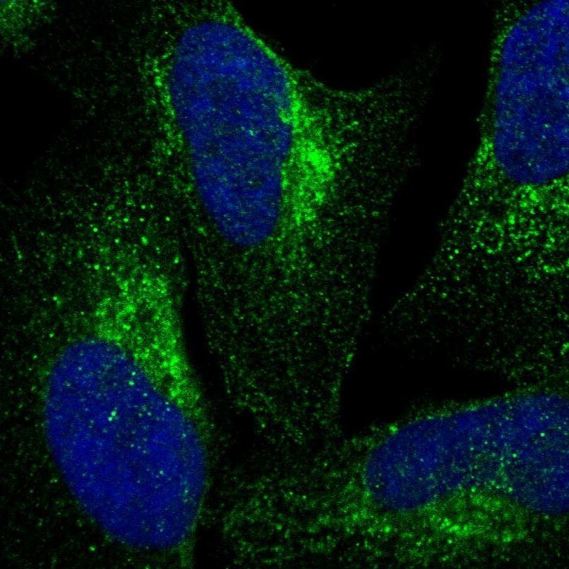
- Immunofluorescent staining of human cell line U-2 OS shows localization to cytosol & the Golgi apparatus.
- Sample type
- Human
Supportive validation
- Submitted by
- Atlas Antibodies (provider)
- Enhanced method
- Orthogonal validation
- Main image

- Experimental details
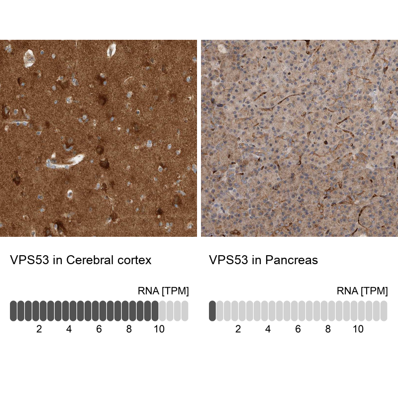
- Immunohistochemistry analysis in human cerebral cortex and pancreas tissues using Anti-VPS53 antibody. Corresponding VPS53 RNA-seq data are presented for the same tissues.
- Sample type
- Human
- Protocol
- Protocol