 Explore
 Validate
 Learn
Learn Western blot
 Immunohistochemistry
Antibody data
- Antibody Data
- Antigen structure
- References [1]
- Comments [0]
- Validations
- Immunohistochemistry [1]
- Flow cytometry [3]
Submit
Validation data
Reference
Comment
Report error
- Product number
- PA5-71494 - Provider product page

- Provider
- Invitrogen Antibodies
- Product name
- IGF2 Polyclonal Antibody
- Antibody type
- Polyclonal
- Antigen
- Synthetic peptide
- Reactivity
- Human, Mouse
- Host
- Rabbit
- Isotype
- IgG
- Vial size
- 200 μL
- Concentration
- 0.5 mg/mL
- Storage
- Store at 4°C short term. For long term storage, store at -20°C, avoiding freeze/thaw cycles.
Submitted references Imprinted Maternally Expressed microRNAs Antagonize Paternally Driven Gene Programs in Neurons.
Whipple AJ, Breton-Provencher V, Jacobs HN, Chitta UK, Sur M, Sharp PA
Molecular cell 2020 Apr 2;78(1):85-95.e8
Molecular cell 2020 Apr 2;78(1):85-95.e8
No comments: Submit comment
Supportive validation
- Submitted by
- Invitrogen Antibodies (provider)
- Main image
- Experimental details
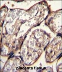
- Immunohistochemistry analysis of IGF2 in formalin fixed and paraffin embedded human placenta tissue. Samples were incubated with IGF2 polyclonal antibody (Product # PA5-71494) followed by peroxidase conjugation of the secondary antibody and DAB staining. This data demonstrates the use of this antibody for immunohistochemistry. Clinical relevance has not been evaluated.

Supportive validation
- Submitted by
- Invitrogen Antibodies (provider)
- Main image

- Experimental details
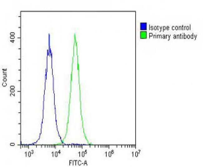
- Overlay histogram showing Hela cells stained with PA5-71494 (green line). The cells were fixed with 2% paraformaldehyde (10 min) and then permeabilized with 90% methanol for 10 min. The cells were then icubated in 2% bovine serum albumin to block non-specific protein-protein interactions followed by the antibody (PA5-71494, 1:25 dilution) for 60 min at 37°C. The secondary antibody used was Goat-Anti-Rabbit IgG, DyLight® 488 Conjugated Highly Cross-Adsorbed(OH191631) at 1/200 dilution for 40 min at 37°C. Isotype control antibody (blue line) was rabbit IgG (1µg/1x10^6 cells) used under the same conditions. Acquisition of >10,000 events was performed.
- Submitted by
- Invitrogen Antibodies (provider)
- Main image

- Experimental details
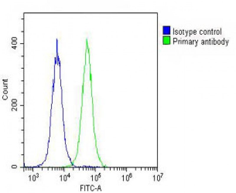
- Flow cytometry analysis of IGF2 in HeLa cells (green line). The cells were fixed with 2% paraformaldhyde (10 min) and permeabilized with 90% methanol (10 min). The sample was then incubated with 2% BSA followed by a IGF2 polyclonal antibody (Product # PA5-71494) using a dilution of 1:25 for 60 min at 37°C. The secondary antibody used was goat-anti-rabbit IgG, DyLight 488 with a dilution of 1:200 for 40 min at 37°C. Isotype control antibody (blue line) was rabbit IgG (1 µg/1x10^6 cells) used under the same conditions with an acquisition of >10, 000 events.
- Submitted by
- Invitrogen Antibodies (provider)
- Main image
- Experimental details
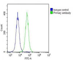
- Flow cytometry of (overlay histogram) of IGF2 in Hela cells (green line). Samples were incubated with IGF2 polyclonal antibody (Product # PA5-71494) using a dilution of 1:25 dilution for 60 min at 37°C followed by Goat-Anti-Rabbit IgG, DyLight® 488 Conjugated Highly Cross-Adsorbed at 1:200 dilution for 40 min at 37°C. The cells were fixed with 2% paraformaldehyde (10 min) and then permeabilized with 90% methanol for 10 min. The cells were then incubated in 2% bovine serum albumin to block non-specific protein-protein interactions followed by the primary antibody. Isotype control antibody (blue line) was rabbit IgG (1 μg/1x10^6 cells) used under the same conditions. Acquisition of >10, 000 events was performed.
