Explore
 Validate
 Learn
Learn Western blot
 Immunoprecipitation
Antibody data
- Antibody Data
- Antigen structure
- References [0]
- Comments [0]
- Validations
- Western blot [6]
- Immunocytochemistry [2]
- Immunohistochemistry [2]
Submit
Validation data
Reference
Comment
Report error
- Product number
- PA5-34786 - Provider product page

- Provider
- Invitrogen Antibodies
- Product name
- Calreticulin Polyclonal Antibody
- Antibody type
- Polyclonal
- Antigen
- Recombinant protein fragment
- Description
- Recommended positive controls: 293T, A431, HeLa, HepG2, A375, Neuro 2A, C8D30, NIH-3T3, Raw264.7, C2C12, PC-12, Rat2. Predicted reactivity: Mouse (94%), Rat (94%), Zebrafish (82%), Xenopus laevis (83%), Pig (94%), Rabbit (95%), Rhesus Monkey (100%), Bovine (95%). Store product as a concentrated solution. Centrifuge briefly prior to opening the vial.
- Reactivity
- Human, Mouse, Rat
- Host
- Rabbit
- Isotype
- IgG
- Vial size
- 100 µL
- Concentration
- 0.2 mg/mL
- Storage
- Store at 4°C short term. For long term storage, store at -20°C, avoiding freeze/thaw cycles.
No comments: Submit comment
Supportive validation
- Submitted by
- Invitrogen Antibodies (provider)
- Main image
- Experimental details
- Western blot analysis of Calreticulin using Various whole cell extracts (30 µg). Samples were loaded onto a 10% SDS-PAGE gel and probed with a Calreticulin polyclonal antibody (Product # PA5-34786) at a dilution of 1:1000.

- Submitted by
- Invitrogen Antibodies (provider)
- Main image

- Experimental details
- Western Blot analysis of Calreticulin was performed by separating 30 µg of various whole cell extracts by 10% SDS-PAGE. Proteins were transferred to a membrane and probed with a Calreticulin Polyclonal Antibody (Product # PA5-34786) at a dilution of 1:1000 and a HRP-conjugated anti-rabbit IgG secondary antibody.

- Submitted by
- Invitrogen Antibodies (provider)
- Main image

- Experimental details
- Western Blot using Calreticulin Polyclonal Antibody (Product # PA5-34786). Various whole cell extracts (30 µg) were separated by 10% SDS-PAGE, and the membrane was blotted with Calreticulin Polyclonal Antibody (Product # PA5-34786) diluted at 1:1,000. The HRP-conjugated anti-rabbit IgG antibody was used to detect the primary antibody.

- Submitted by
- Invitrogen Antibodies (provider)
- Main image

- Experimental details
- Western Blot analysis of Calreticulin was performed by separating 30 µg of various whole cell extracts by 10% SDS-PAGE. Proteins were transferred to a membrane and probed with a Calreticulin Polyclonal Antibody (Product # PA5-34786) at a dilution of 1:1000 and a HRP-conjugated anti-rabbit IgG secondary antibody.

- Submitted by
- Invitrogen Antibodies (provider)
- Main image

- Experimental details
- Knockdown of Calreticulin was achieved by transfecting HeLa cells with Calreticulin specific siRNAs (Silencer® select Product # s114). Western blot analysis (Fig. a) was performed using whole cell extracts from the Calreticulin knockdown cells (lane 3), non-specific scrambled siRNA transfected cells (lane 2) and untransfected cells (lane 1). The blots were probed with Calreticulin Polyclonal Antibody (Product # PA5-34786, 1:2000 dilution) and Goat anti-Rabbit IgG (H+L) Superclonal™ Secondary Antibody, HRP conjugate (Product # A27036, 0.25µg/ml, 1:4000 dilution). Densitometric analysis of this western blot is shown in histogram (Fig. b). Decrease in signal upon siRNA mediated knock down confirms that antibody is specific to Calreticulin.

- Submitted by
- Invitrogen Antibodies (provider)
- Main image

- Experimental details
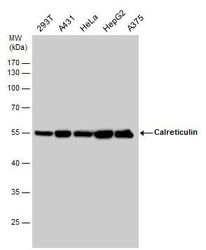
- Western blot analysis was performed on whole cell extract (30 µg lysate) of RAW 264.7 (Lane 1), SHSY5Y (Lane 2), HeLa (Lane 3), A431 (Lane 4), SKOV3 (Lane 5), IM9 (Lane 6), Jurkat (Lane 7), K562 (Lane 8), and MCF7 (Lane 9). The blot was probed with Anti-Calreticulin Polyclonal Antibody (Product # PA5-34786, 1:1000 dilution) and detected by chemiluminescence using Goat anti-Rabbit IgG (H+L) Superclonal™ Secondary Antibody, HRP conjugate (Product # A27036, 0.25 µg/ml, 1:4000 dilution). A 55 kDa band corresponding to Calreticulin was observed in all cell lines tested.

Supportive validation
- Submitted by
- Invitrogen Antibodies (provider)
- Main image

- Experimental details
- Immunofluorescent analysis of Calreticulin showing staining in the endoplasmic reticulum of Jurkat cells. Jurkat cells were fixed in 4% paraformaldehyde at RT for 15 min and stained using a Calreticulin polyclonal antibody (Product # PA5-34786) diluted at 1:500. Blue: Hoechst 33342 staining. Scale bar = 10µm.
- Submitted by
- Invitrogen Antibodies (provider)
- Main image

- Experimental details
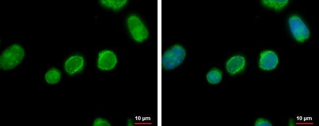
- Calreticulin Polyclonal Antibody detects Calreticulin protein at endoplasmic reticulum by immunofluorescent analysis. Sample: HeLa cells were fixed in ice-cold MeOH for 5 min. Green: Calreticulin stained by Calreticulin Polyclonal Antibody (Product # PA5-34786) diluted at 1:500.

Supportive validation
- Submitted by
- Invitrogen Antibodies (provider)
- Main image

- Experimental details
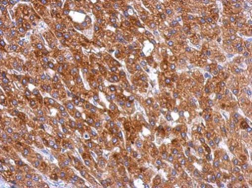
- Immunohistochemical analysis of paraffin-embedded human hepatoma, using Calreticulin (Product # PA5-34786) antibody at 1:500 dilution. Antigen Retrieval: EDTA based buffer, pH 8.0, 15 min.
- Submitted by
- Invitrogen Antibodies (provider)
- Main image
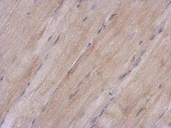
- Experimental details
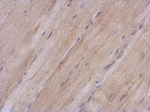
- Immunohistochemical analysis of paraffin-embedded mouse muscle, using Calreticulin (Product # PA5-34786) antibody at 1:500 dilution. Antigen Retrieval: EDTA based buffer, pH 8.0, 15 min.