Explore
 Validate
 Learn
Learn Western blot
 Immunocytochemistry
Antibody data
- Antibody Data
- Antigen structure
- References [4]
- Comments [0]
- Validations
- Immunocytochemistry [7]
- Immunohistochemistry [1]
- Flow cytometry [2]
- Other assay [1]
Submit
Validation data
Reference
Comment
Report error
- Product number
- MA5-15382 - Provider product page

- Provider
- Invitrogen Antibodies
- Product name
- Calreticulin Monoclonal Antibody (1G6A7)
- Antibody type
- Monoclonal
- Antigen
- Synthetic peptide
- Description
- MA5-15382 targets Calreticulin in IF, IHC, Flow and WB applications and shows reactivity with Human, non-Human primate, and mouse samples. The MA5-15382 immunogen is synthetic peptide corresponding to aa (EEEDVPGQAKDELC) of human Calreticulin, conjugated to KLH. MA5-15382 detects Calreticulin which has a predicted molecular weight of approximately 48kDa.
- Reactivity
- Human, Mouse
- Host
- Mouse
- Isotype
- IgG
- Antibody clone number
- 1G6A7
- Vial size
- 100 μg
- Concentration
- 1 mg/mL
- Storage
- Store at 4°C short term. For long term storage, store at -20°C, avoiding freeze/thaw cycles.
Submitted references Expression in retinal neurons of fukutin and FKRP, the protein products of two dystroglycanopathy-causative genes.
Transcriptomic and proteomic host response to Aspergillus fumigatus conidia in an air-liquid interface model of human bronchial epithelium.
Serum calreticulin is a negative biomarker in patients with Alzheimer's disease.
Platelet protein disulfide isomerase is localized in the dense tubular system and does not become surface expressed after activation.
Haro C, Uribe ML, Quereda C, Cruces J, Martín-Nieto J
Molecular vision 2018;24:43-58
Molecular vision 2018;24:43-58
Transcriptomic and proteomic host response to Aspergillus fumigatus conidia in an air-liquid interface model of human bronchial epithelium.
Toor A, Culibrk L, Singhera GK, Moon KM, Prudova A, Foster LJ, Moore MM, Dorscheid DR, Tebbutt SJ
PloS one 2018;13(12):e0209652
PloS one 2018;13(12):e0209652
Serum calreticulin is a negative biomarker in patients with Alzheimer's disease.
Lin Q, Cao Y, Gao J
International journal of molecular sciences 2014 Nov 25;15(12):21740-53
International journal of molecular sciences 2014 Nov 25;15(12):21740-53
Platelet protein disulfide isomerase is localized in the dense tubular system and does not become surface expressed after activation.
van Nispen Tot Pannerden HE, van Dijk SM, Du V, Heijnen HF
Blood 2009 Nov 19;114(21):4738-40
Blood 2009 Nov 19;114(21):4738-40
No comments: Submit comment
Supportive validation
- Submitted by
- Invitrogen Antibodies (provider)
- Main image
- Experimental details
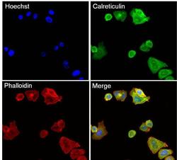
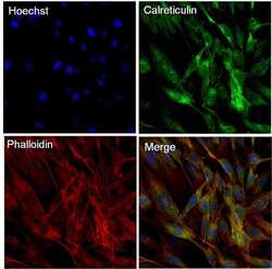
- Immunofluorescent analysis of Calreticulin (green) in SK-BR-3 cells. The cells were fixed with 4% paraformaldehyde in PBS for 15 minutes at room temperature, permeabilized with 0.1% Triton X-100 for 15 minutes, and blocked with 3% BSA for 30 minutes at room temperature. Cells were stained with a Calreticulin mouse monoclonal antibody (Product # MA5-15382) at a concentration of 10 µg/mL in blocking buffer for 1 hour at room temperature, and then incubated with a Goat anti-Mouse IgG (H+L) Secondary Antibody, Alexa Fluor Plus 488 conjugate (Product # A32731) at a dilution of 1:500 for at least 30 minutes at a room temperature in the dark (green). F-actin (red) was stained by Dylight 554 Phalloidin (Product # 21834) and nuclei (blue) were stained with Hoechst 33342 (Product # 62249). Images were taken on a Thermo Scientific ToxInsight Instrument at 20X magnification.
- Submitted by
- Invitrogen Antibodies (provider)
- Main image

- Experimental details
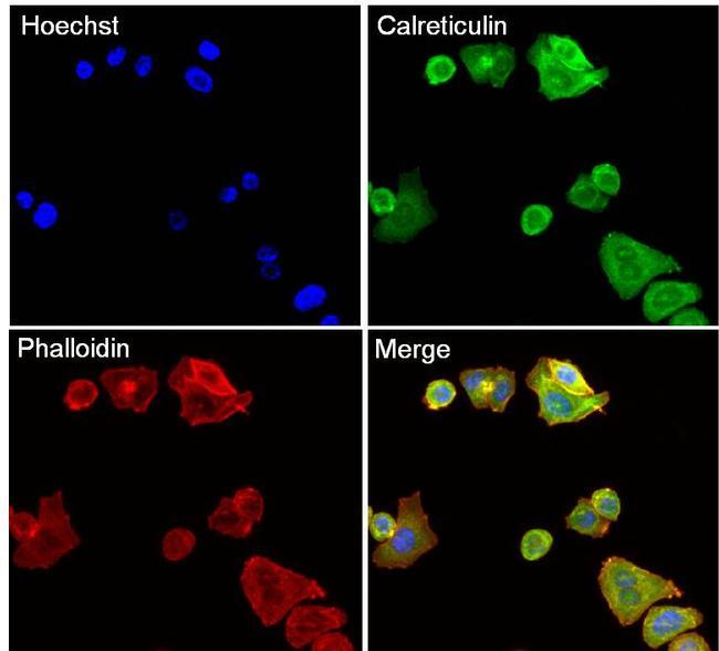
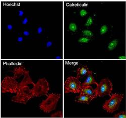
- Immunofluorescent analysis of Calreticulin (green) in A549 cells. The cells were fixed with 4% paraformaldehyde in PBS for 15 minutes at room temperature, permeabilized with 0.1% Triton X-100 for 15 minutes, and blocked with 3% BSA for 30 minutes at room temperature. Cells were stained with a Calreticulin mouse monoclonal antibody (Product # MA5-15382) at a concentration of 10 µg/mL in blocking buffer for 1 hour at room temperature, and then incubated with a Goat anti-Mouse IgG (H+L) Secondary Antibody, Alexa Fluor Plus 488 conjugate (Product # A32731) at a dilution of 1:500 for at least 30 minutes at a room temperature in the dark (green). F-actin (red) was stained by Dylight 554 Phalloidin (Product # 21834) and nuclei (blue) were stained with Hoechst 33342 (Product # 62249). Images were taken on a Thermo Scientific ToxInsight Instrument at 20X magnification.
- Submitted by
- Invitrogen Antibodies (provider)
- Main image

- Experimental details
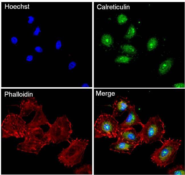
- Immunofluorescent analysis of Calreticulin (green) in SK-BR-3 cells. The cells were fixed with 4% paraformaldehyde in PBS for 15 minutes at room temperature, permeabilized with 0.1% Triton X-100 for 15 minutes, and blocked with 3% BSA for 30 minutes at room temperature. Cells were stained with a Calreticulin mouse monoclonal antibody (Product # MA5-15382) at a concentration of 10 µg/mL in blocking buffer for 1 hour at room temperature, and then incubated with a Goat anti-Mouse IgG (H+L) Secondary Antibody, Alexa Fluor Plus 488 conjugate (Product # A32731) at a dilution of 1:500 for at least 30 minutes at a room temperature in the dark (green). F-actin (red) was stained by Dylight 554 Phalloidin (Product # 21834) and nuclei (blue) were stained with Hoechst 33342 (Product # 62249). Images were taken on a Thermo Scientific ToxInsight Instrument at 20X magnification.

- Submitted by
- Invitrogen Antibodies (provider)
- Main image
- Experimental details
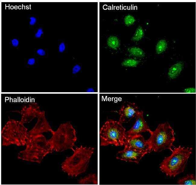
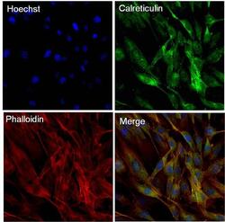
- Immunofluorescent analysis of Calreticulin (green) in NIH3T3 cells. The cells were fixed with 4% paraformaldehyde in PBS for 15 minutes at room temperature, permeabilized with 0.1% Triton X-100 for 15 minutes, and blocked with 3% BSA for 30 minutes at room temperature. Cells were stained with a Calreticulin mouse monoclonal antibody (Product # MA5-15382) at a concentration of 10 µg/mL in blocking buffer for 1 hour at room temperature, and then incubated with a Goat anti-Mouse IgG (H+L) Secondary Antibody, Alexa Fluor Plus 488 conjugate (Product # A32731) at a dilution of 1:500 for at least 30 minutes at a room temperature in the dark (green). F-actin (red) was stained by Dylight 554 Phalloidin (Product # 21834) and nuclei (blue) were stained with Hoechst 33342 (Product # 62249). Images were taken on a Thermo Scientific ToxInsight Instrument at 20X magnification.

- Submitted by
- Invitrogen Antibodies (provider)
- Main image
- Experimental details
- Immunofluorescent analysis of Calreticulin (green) in A549 cells. The cells were fixed with 4% paraformaldehyde in PBS for 15 minutes at room temperature, permeabilized with 0.1% Triton X-100 for 15 minutes, and blocked with 3% BSA for 30 minutes at room temperature. Cells were stained with a Calreticulin mouse monoclonal antibody (Product # MA5-15382) at a concentration of 10 µg/mL in blocking buffer for 1 hour at room temperature, and then incubated with a Goat anti-Mouse IgG (H+L) Secondary Antibody, Alexa Fluor Plus 488 conjugate (Product # A32731) at a dilution of 1:500 for at least 30 minutes at a room temperature in the dark (green). F-actin (red) was stained by Dylight 554 Phalloidin (Product # 21834) and nuclei (blue) were stained with Hoechst 33342 (Product # 62249). Images were taken on a Thermo Scientific ToxInsight Instrument at 20X magnification.
- Submitted by
- Invitrogen Antibodies (provider)
- Main image

- Experimental details
- Immunofluorescent analysis of Calreticulin (green) in SK-BR-3 cells. The cells were fixed with 4% paraformaldehyde in PBS for 15 minutes at room temperature, permeabilized with 0.1% Triton X-100 for 15 minutes, and blocked with 3% BSA for 30 minutes at room temperature. Cells were stained with a Calreticulin mouse monoclonal antibody (Product # MA5-15382) at a concentration of 10 µg/mL in blocking buffer for 1 hour at room temperature, and then incubated with a Goat anti-Mouse IgG (H+L) Secondary Antibody, Alexa Fluor Plus 488 conjugate (Product # A32731) at a dilution of 1:500 for at least 30 minutes at a room temperature in the dark (green). F-actin (red) was stained by Dylight 554 Phalloidin (Product # 21834) and nuclei (blue) were stained with Hoechst 33342 (Product # 62249). Images were taken on a Thermo Scientific ToxInsight Instrument at 20X magnification.

- Submitted by
- Invitrogen Antibodies (provider)
- Main image
- Experimental details
- Immunofluorescent analysis of Calreticulin (green) in NIH3T3 cells. The cells were fixed with 4% paraformaldehyde in PBS for 15 minutes at room temperature, permeabilized with 0.1% Triton X-100 for 15 minutes, and blocked with 3% BSA for 30 minutes at room temperature. Cells were stained with a Calreticulin mouse monoclonal antibody (Product # MA5-15382) at a concentration of 10 µg/mL in blocking buffer for 1 hour at room temperature, and then incubated with a Goat anti-Mouse IgG (H+L) Secondary Antibody, Alexa Fluor Plus 488 conjugate (Product # A32731) at a dilution of 1:500 for at least 30 minutes at a room temperature in the dark (green). F-actin (red) was stained by Dylight 554 Phalloidin (Product # 21834) and nuclei (blue) were stained with Hoechst 33342 (Product # 62249). Images were taken on a Thermo Scientific ToxInsight Instrument at 20X magnification.

Supportive validation
- Submitted by
- Invitrogen Antibodies (provider)
- Main image
- Experimental details
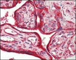
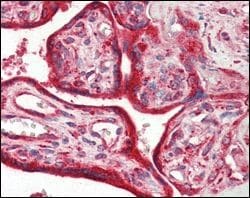
- Immunohistochemical analysis of paraffin-embedded human placenta tissues using Calreticulin monoclonal antibody (Product # MA5-15382).
Supportive validation
- Submitted by
- Invitrogen Antibodies (provider)
- Main image

- Experimental details
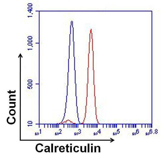
- Flow cytometry analysis of Calreticulin was done on HeLa cells. The cells were fixed, permeabilized and stained with a Calreticulin mouse monoclonal antibody (Product # MA5-15382, red histogram) or Mouse IgG2a isotype control (Product # MA1-10418, blue histogram) at a concentration of 2µg/mL. After incubation of the primary antibody on ice for an hour, the cells were stained with a Goat anti-Mouse IgG (H+L) Secondary Antibody, Alexa Fluor Plus 680 conjugate (Product # A32734) at a dilution of 1:50 for at least 30 minutes on ice. A representative 10,000 cells were acquired for each sample.
- Submitted by
- Invitrogen Antibodies (provider)
- Main image
- Experimental details
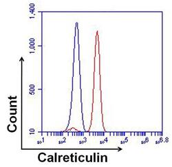
- Flow cytometry analysis of Calreticulin was done on HeLa cells. The cells were fixed, permeabilized and stained with a Calreticulin mouse monoclonal antibody (Product # MA5-15382, red histogram) or Mouse IgG2a isotype control (Product # MA1-10418, blue histogram) at a concentration of 2µg/mL. After incubation of the primary antibody on ice for an hour, the cells were stained with a Goat anti-Mouse IgG (H+L) Secondary Antibody, Alexa Fluor Plus 680 conjugate (Product # A32734) at a dilution of 1:50 for at least 30 minutes on ice. A representative 10,000 cells were acquired for each sample.

Supportive validation
- Submitted by
- Invitrogen Antibodies (provider)
- Main image

- Experimental details
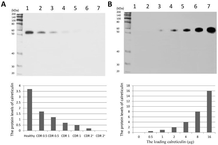
- Figure 3 Western Blot analysis of serum calreticulin of AD patients and healthy subjects. ( A ) serum levels of calreticulin in AD patients and healthy subjects. Lane 1, a healthy control; Lanes 2-7, AD patients and ( B ) lanes 1-7, 0, 0.5, 1, 2, 4, 8 and 16 ug of calreticulin was loaded in respectively. A band of approximately 55-kDa protein could be detected (predicted molecular weight: 48 kDa).