Explore
 Validate
 Learn
Learn Western blot
Antibody data
- Antibody Data
- Antigen structure
- References [12]
- Comments [0]
- Validations
- Western blot [3]
- Other assay [9]
Submit
Validation data
Reference
Comment
Report error
- Product number
- PA1-902A - Provider product page

- Provider
- Invitrogen Antibodies
- Product name
- Calreticulin Polyclonal Antibody
- Antibody type
- Polyclonal
- Antigen
- Synthetic peptide
- Description
- PA1-902A detects calreticulin from canine, hamster, human, mouse and rat tissues. PA1-902A has been successfully used in Western blot, immunocytochemistry and immunofluorescene procedures. By Western blot, this antibody detects an ~60-66 kDa protein representing calreticulin. PA1-902A immunizing peptide corresponds to amino acid residues 24-43 from mouse calreticulin. PA1-902A immunizing peptide (Cat. # PEP-018) is available for use in neutralization and control experiments.
- Reactivity
- Human, Mouse, Rat, Canine, Hamster
- Host
- Chicken/Avian
- Isotype
- IgY
- Vial size
- 100 μL
- Concentration
- 1.1 mg/mL
- Storage
- -20°C, Avoid Freeze/Thaw Cycles
Submitted references Adaptive changes of telocytes in the urinary bladder of patients affected by neurogenic detrusor overactivity.
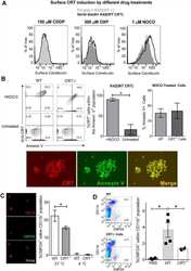
Calreticulin Release at an Early Stage of Death Modulates the Clearance by Macrophages of Apoptotic Cells.
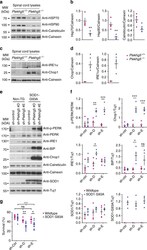
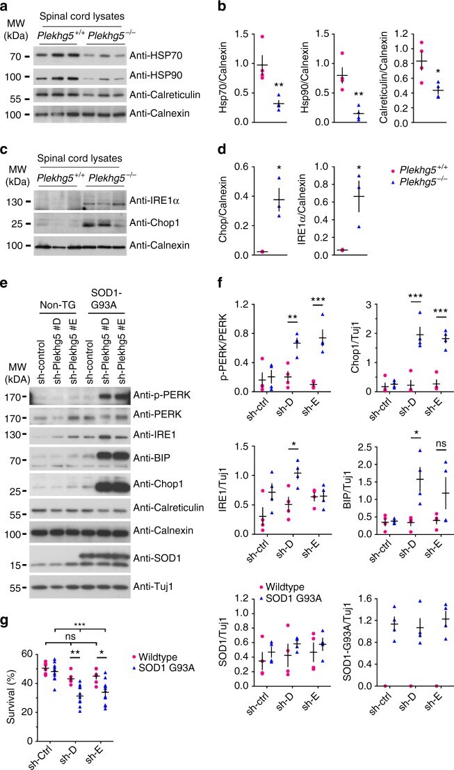
Plekhg5-regulated autophagy of synaptic vesicles reveals a pathogenic mechanism in motoneuron disease.
The C-Terminal Acidic Region of Calreticulin Mediates Phosphatidylserine Binding and Apoptotic Cell Phagocytosis.
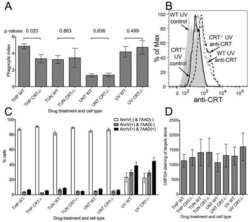
Concurrent MEK and autophagy inhibition is required to restore cell death associated danger-signalling in Vemurafenib-resistant melanoma cells.
Inner and outer portions of colonic circular muscle: ultrastructural and immunohistochemical changes in rat chronically treated with otilonium bromide.
Relative contribution of c1q and apoptotic cell-surface calreticulin to macrophage phagocytosis.
Telocytes subtypes in human urinary bladder.
Chronic ethanol consumption in mice alters hepatocyte lipid droplet properties.
Endoplasmic reticulum calcium depletion impacts chaperone secretion, innate immunity, and phagocytic uptake of cells.
The WFS1 gene, responsible for low frequency sensorineural hearing loss and Wolfram syndrome, is expressed in a variety of inner ear cells.
Cytoplasmic domain of herpes simplex virus gE causes accumulation in the trans-Golgi network, a site of virus envelopment and sorting of virions to cell junctions.
Traini C, Fausssone-Pellegrini MS, Guasti D, Del Popolo G, Frizzi J, Serni S, Vannucchi MG
Journal of cellular and molecular medicine 2018 Jan;22(1):195-206
Journal of cellular and molecular medicine 2018 Jan;22(1):195-206
Calreticulin Release at an Early Stage of Death Modulates the Clearance by Macrophages of Apoptotic Cells.
Osman R, Tacnet-Delorme P, Kleman JP, Millet A, Frachet P
Frontiers in immunology 2017;8:1034
Frontiers in immunology 2017;8:1034
Plekhg5-regulated autophagy of synaptic vesicles reveals a pathogenic mechanism in motoneuron disease.
Lüningschrör P, Binotti B, Dombert B, Heimann P, Perez-Lara A, Slotta C, Thau-Habermann N, R von Collenberg C, Karl F, Damme M, Horowitz A, Maystadt I, Füchtbauer A, Füchtbauer EM, Jablonka S, Blum R, Üçeyler N, Petri S, Kaltschmidt B, Jahn R, Kaltschmidt C, Sendtner M
Nature communications 2017 Oct 30;8(1):678
Nature communications 2017 Oct 30;8(1):678
The C-Terminal Acidic Region of Calreticulin Mediates Phosphatidylserine Binding and Apoptotic Cell Phagocytosis.
Wijeyesakere SJ, Bedi SK, Huynh D, Raghavan M
Journal of immunology (Baltimore, Md. : 1950) 2016 May 1;196(9):3896-3909
Journal of immunology (Baltimore, Md. : 1950) 2016 May 1;196(9):3896-3909
Concurrent MEK and autophagy inhibition is required to restore cell death associated danger-signalling in Vemurafenib-resistant melanoma cells.
Martin S, Dudek-Perić AM, Maes H, Garg AD, Gabrysiak M, Demirsoy S, Swinnen JV, Agostinis P
Biochemical pharmacology 2015 Feb 1;93(3):290-304
Biochemical pharmacology 2015 Feb 1;93(3):290-304
Inner and outer portions of colonic circular muscle: ultrastructural and immunohistochemical changes in rat chronically treated with otilonium bromide.
Traini C, Faussone-Pellegrini MS, Evangelista S, Mazzaferro K, Cipriani G, Santicioli P, Vannucchi MG
PloS one 2014;9(8):e103237
PloS one 2014;9(8):e103237
Relative contribution of c1q and apoptotic cell-surface calreticulin to macrophage phagocytosis.
Verneret M, Tacnet-Delorme P, Osman R, Awad R, Grichine A, Kleman JP, Frachet P
Journal of innate immunity 2014;6(4):426-34
Journal of innate immunity 2014;6(4):426-34
Telocytes subtypes in human urinary bladder.
Vannucchi MG, Traini C, Guasti D, Del Popolo G, Faussone-Pellegrini MS
Journal of cellular and molecular medicine 2014 Oct;18(10):2000-8
Journal of cellular and molecular medicine 2014 Oct;18(10):2000-8
Chronic ethanol consumption in mice alters hepatocyte lipid droplet properties.
Orlicky DJ, Roede JR, Bales E, Greenwood C, Greenberg A, Petersen D, McManaman JL
Alcoholism, clinical and experimental research 2011 Jun;35(6):1020-33
Alcoholism, clinical and experimental research 2011 Jun;35(6):1020-33
Endoplasmic reticulum calcium depletion impacts chaperone secretion, innate immunity, and phagocytic uptake of cells.
Peters LR, Raghavan M
Journal of immunology (Baltimore, Md. : 1950) 2011 Jul 15;187(2):919-31
Journal of immunology (Baltimore, Md. : 1950) 2011 Jul 15;187(2):919-31
The WFS1 gene, responsible for low frequency sensorineural hearing loss and Wolfram syndrome, is expressed in a variety of inner ear cells.
Cryns K, Thys S, Van Laer L, Oka Y, Pfister M, Van Nassauw L, Smith RJ, Timmermans JP, Van Camp G
Histochemistry and cell biology 2003 Mar;119(3):247-56
Histochemistry and cell biology 2003 Mar;119(3):247-56
Cytoplasmic domain of herpes simplex virus gE causes accumulation in the trans-Golgi network, a site of virus envelopment and sorting of virions to cell junctions.
McMillan TN, Johnson DC
Journal of virology 2001 Feb;75(4):1928-40
Journal of virology 2001 Feb;75(4):1928-40
No comments: Submit comment
Supportive validation
- Submitted by
- Invitrogen Antibodies (provider)
- Main image

- Experimental details
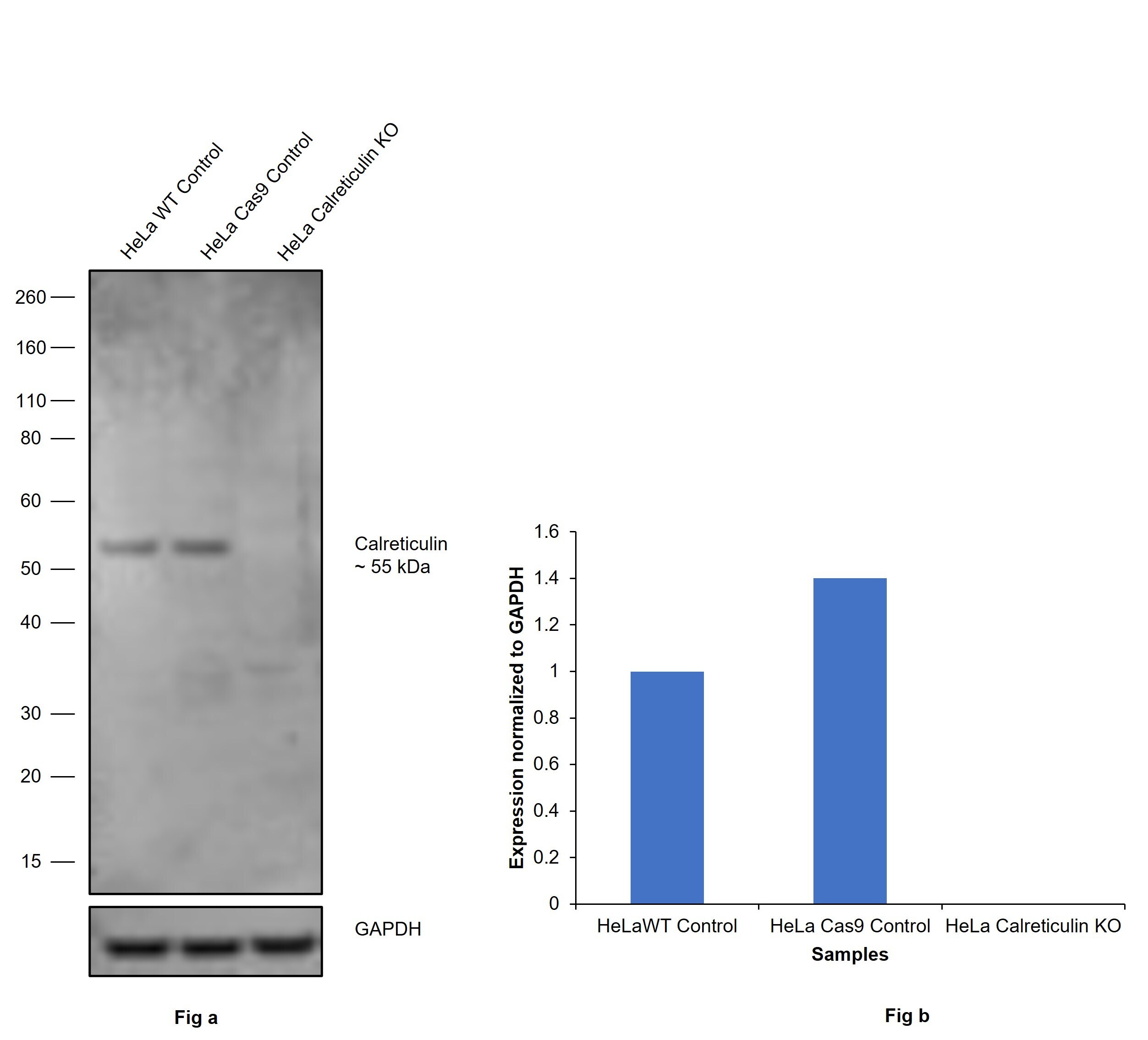
- Knockout of Calreticulin was achieved by CRISPR-Cas9 genome editing using LentiArray™ Lentiviral sgRNA (Product # A32042, Assay ID CRISPR1007602_LV) and LentiArray Cas9 Lentivirus (Product # A32064). Western blot analysis of Calreticulin was performed by loading 30 µg of HeLa wild type (Lane 1), HeLa Cas9 (Lane 2) and HeLa Calreticulin KO (Lane 3) whole cell extracts. The samples were electrophoresed using NuPAGE™ Novex™ 4-12% Bis-Tris Protein Gel (Product # NP0322BOX). Resolved proteins were then transferred onto a nitrocellulose membrane (Product # IB23001) by iBlot® 2 Dry Blotting System (Product # IB21001). The blot was probed with Calreticulin Polyclonal Antibody (Product # PA1-902A, 1:1000 dilution) and Goat anti-Chicken IgY (H+L) Secondary Antibody, HRP (Product # A16054, 1:10,000 dilution) using the iBright™ FL1500 (Product # A44115). Chemiluminescent detection was performed usingSuperSignal™ West Dura Extended Duration Substrate (Product # 34076). Loss of signal upon CRISPR mediated knockout (KO) using the LentiArray™ CRISPR product line confirms that antibody is specific to Calreticulin.
- Submitted by
- Invitrogen Antibodies (provider)
- Main image
- Experimental details
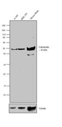
- Western blot analysis was performed on membrane enriched extracts (30 µg lysate) of A-549 (Lane 1), HEK 293 (Lane 2) and Mouse Brain (Lane 3). The blot was probed with Anti-Calreticulin Chicken Polyclonal Antibody (Product # PA1-902A, 1:500-1:2000 dilution) and detected by chemiluminescence using Goat anti-Chicken IgY (H+L) Secondary Antibody, HRP conjugate (Product # A16054, 0.4 µg/mL, 1:2500 dilution). A 48 kDa band corresponding to Calreticulin was observed across the cell lines and tissue tested. Known quantity of protein samples were electrophoresed using Novex® NuPAGE® 10 % Bis-Tris gel (Product # NP0302BOX), XCell SureLock™ Electrophoresis System (Product # EI0002) and Novex® Sharp Pre-Stained Protein Standard (Product # LC5800). Resolved proteins were then transferred onto a nitrocellulose membrane with iBlot® 2 Dry Blotting System (Product # IB21001). The membrane was probed with the relevant primary and secondary Antibody following blocking with 5 % skimmed milk. Chemiluminescent detection was performed using Pierce™ ECL Western Blotting Substrate (Product # 32106).

- Submitted by
- Invitrogen Antibodies (provider)
- Main image
- Experimental details
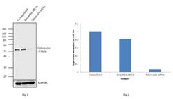
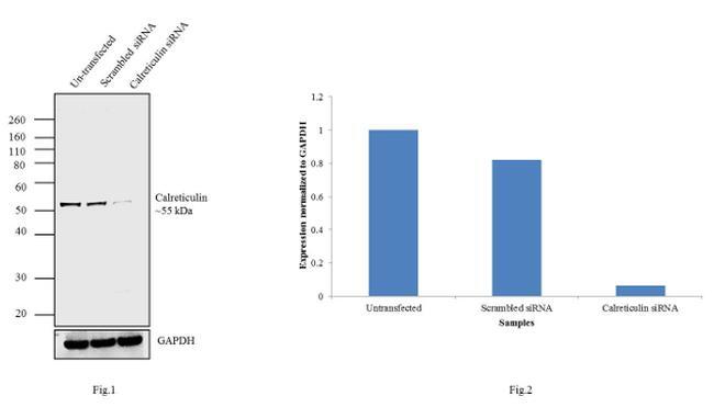
- Knockdown of Calreticulin was achieved by transfecting MCF 7 cells with Calreticulin specific validated siRNAs (Silencer® select Product # s114). Western blot analysis (Fig 1) was performed using whole cell extracts from the Calreticulin knockdown cells (lane 3), non-specific scrambled siRNA transfected cells (lane 2) and untransfected cells (lane 1). The blots were probed with Anti-Calreticulin Antibody, Chicken monoclonal (Product # PA1-902A, 1 µg/mL) and Goat anti-Chicken IgY (H+L) Secondary Antibody, HRP conjugate (Product # A16054, 0.4 µg/mL, 1:4000 dilution). Densitometric analysis of this western blot is shown in histogram (Fig 2). Decrease in signal upon siRNA mediated knock down confirms that antibody is specific to Calreticulin.
Supportive validation
- Submitted by
- Invitrogen Antibodies (provider)
- Main image
- Experimental details
- NULL
- Submitted by
- Invitrogen Antibodies (provider)
- Main image

- Experimental details
- NULL
- Submitted by
- Invitrogen Antibodies (provider)
- Main image
- Experimental details
- NULL

- Submitted by
- Invitrogen Antibodies (provider)
- Main image
- Experimental details
- NULL

- Submitted by
- Invitrogen Antibodies (provider)
- Main image
- Experimental details
- Fig. 9 Plekhg5 depletion in SOD1 G93A motoneurons results in elevated ER-stress. a Expression of HSP70, HSP90, Calreticulin, and Calnexin in spinal cord lysates from three animals per genotype. b Quantification of western blot shown in a (each data point represents expression levels of one animal; unpaired t -test; two-tailed). c Expression of IRE1alpha and Chop1 in spinal cord lysates from three animals per genotype. d Quantification of western blot shown in c (each data point represents expression levels of one animal; unpaired t -test; two-tailed). e SOD1 G93A and non-transgenic motoneurons were depleted of Plekhg5 and several ER-stress markers were examined after 7 days in culture. f Quantification of western blots shown in e (each data point represents one individual experiment; mean +- SEM; unpaired t -test; two-tailed). g Survival of SOD1 G93A motoneurons decreased upon knockdown of Plekhg5 using two independent sh-RNA constructs (each data point represents the % of motoneuron-survival from one individual embryo. At least 50 motoneurons were evaluated from one embryo; mean +- SEM; two-way ANOVA; Bonferroni post-test). Images have been cropped for presentation. Full size images are presented in Supplementary Fig. 7
- Submitted by
- Invitrogen Antibodies (provider)
- Main image
- Experimental details
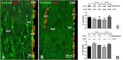
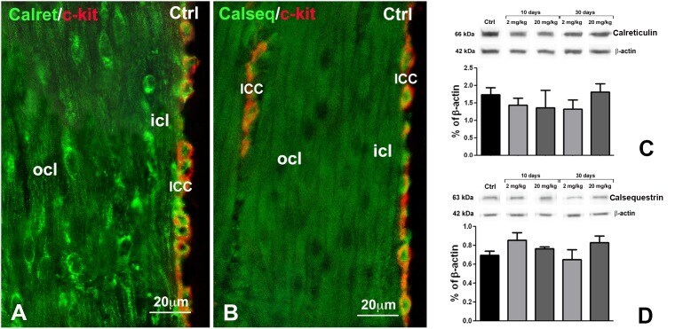
- Figure 5 Immunohistochemistry (IHC) and western blots (WB) for Calret and Calseq. A and B : IHC. Controls. In ( A ), double labelling for Calret (green) and c-kit (red), in ( B ), double labelling for Calseq (green) and c-kit (red). The smooth muscle cells of both ocl and icl are positive, but no differences in the IR intensity are present between the two layers. The ICC are double labelled (green and red). Calret-IR appears as granules mainly distributed at cell periphery, while Calseq-IR is intracytoplasmatic. A , B : Bar = 20 um. C, D : WB. Repre-sentative bands of Calret-IR ( C) and Calseq-IR ( D ) and their quantification in control and treated rats. No significant change was seen at any time of duration of OB treatment for both markers.
- Submitted by
- Invitrogen Antibodies (provider)
- Main image
- Experimental details
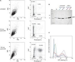
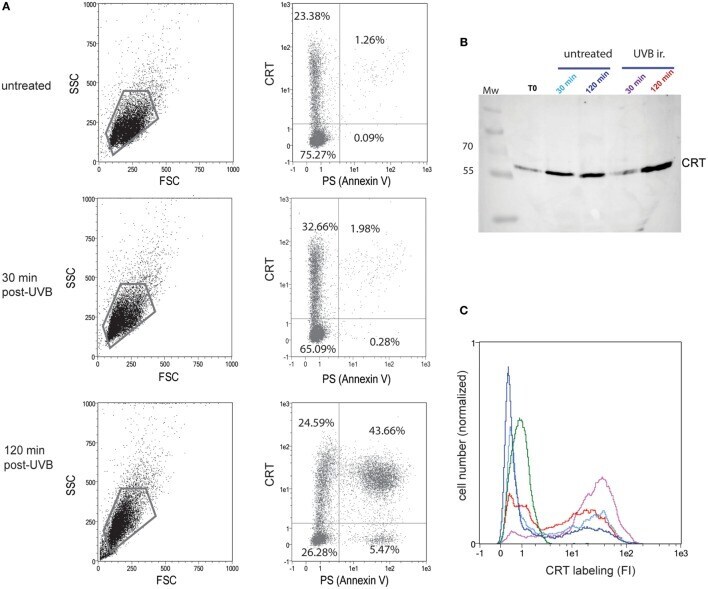
- Figure 1 Calreticulin (CRT) is exposed rapidly after UVB irradiation at the cell surface and released in the medium. (A) Uuntreated or UVB-irradiated JurkaT cells were analyzed by flow cytometry for their phosphatidylserine (PS) (Annexin V-FITC) and CRT (anti-CRT antibody/secondary antibody-Cy3) surface exposure. The region selected for double PS/CRT analysis is shown on SSC/FSC dot plots. Double PS/CRT labeling dot plots: % of cells in quadrants is indicated. (B) CRT detection by western blotting on the medium conditioned by JurkaT cells and collected at different times as mentioned. Each line corresponds to 1 ml of medium conditioned by 2 x 10 6 cells. T0 point corresponds to medium added to untreated cells and immediately recovered. Mw, molecular weight ladders (kilodaltons). (C) In parallel to supernatants recovering, cells used for the analysis shown in B were collected, immediately fixed, and labeled for surface CRT detection by flow cytometry. Ungated populations were analyzed. B and C, The same color code is used for conditioned medium or the corresponding cells: untreated control condition after 30 min of incubation (light blue) or 120 min (dark blue), UVB-irradiated condition after 30 min of incubation (purple) or 120 min (red). An isotype control is shown in green. (A-C) Representative experiments are shown.
- Submitted by
- Invitrogen Antibodies (provider)
- Main image
- Experimental details
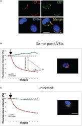
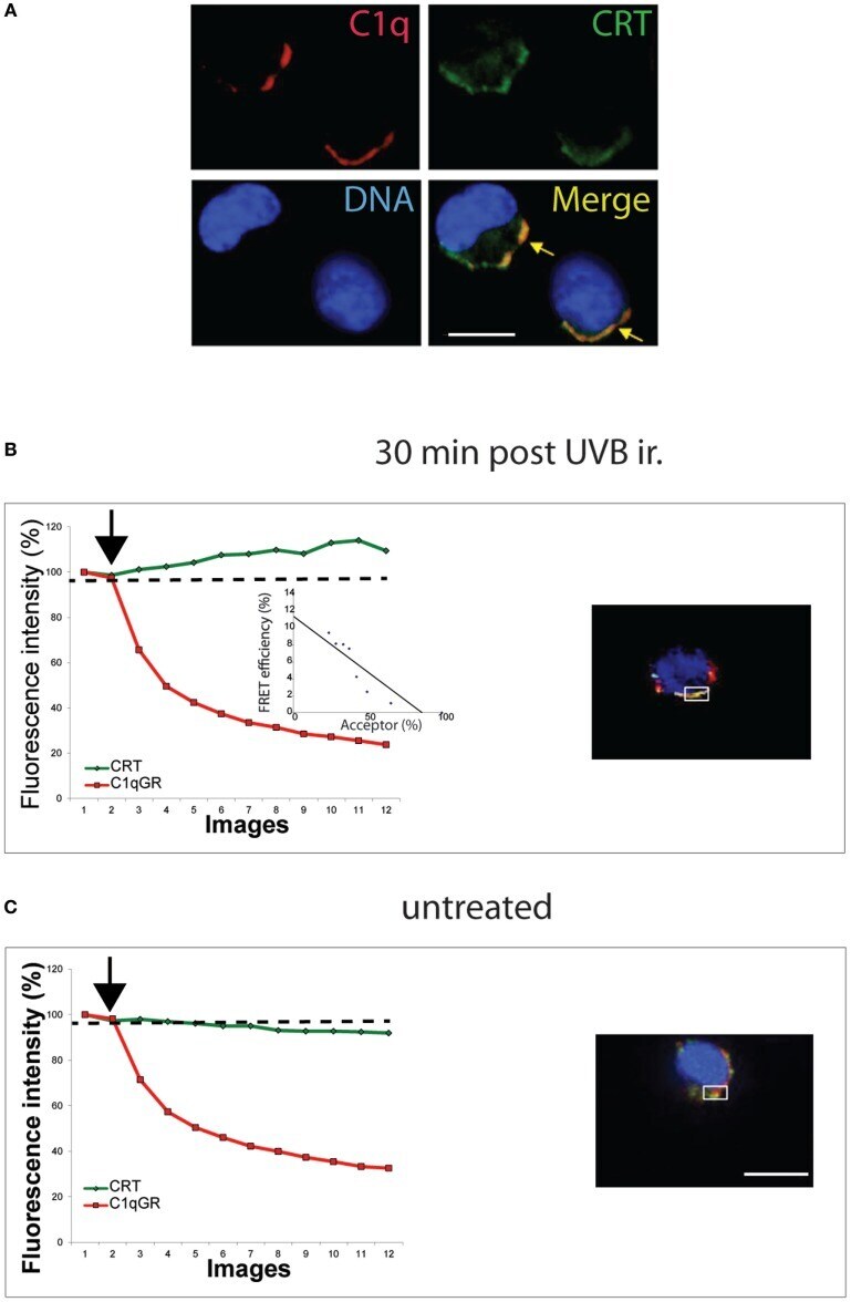
- Figure 2 C1q interacts with ecto-calreticulin very rapidly after UVB irradiation. JurkaT cells incubated with C1q or its globular region were immunolabeled for Calreticulin (CRT) (A488) and C1q or C1qGR (Cy3) as described in Section "" Material and Methods ."" (A) Labeling on cells fixed 30 min after UVB irradiation. (B,C) Fluorescence resonance energy transfer (FRET) efficiency was estimated for UVB-irradiated cell and untreated cell by photobleaching of the acceptor dye (Cy3) on non-permeabilized cells. Regions used for the acceptor photobleaching and FRET analysis are shown. Curves correspond to the normalized fluorescence intensities of both dyes (Cy3 in red and A488 in green) expressed as a percent of the signal measured before the gradual photobleaching started (black arrow). (B) FRET efficiency (percent of acceptor fluorescence intensity increase) is expressed as a function of the percent of the normalized acceptor fluorescence intensity. Bars: 11 um.
- Submitted by
- Invitrogen Antibodies (provider)
- Main image

- Experimental details
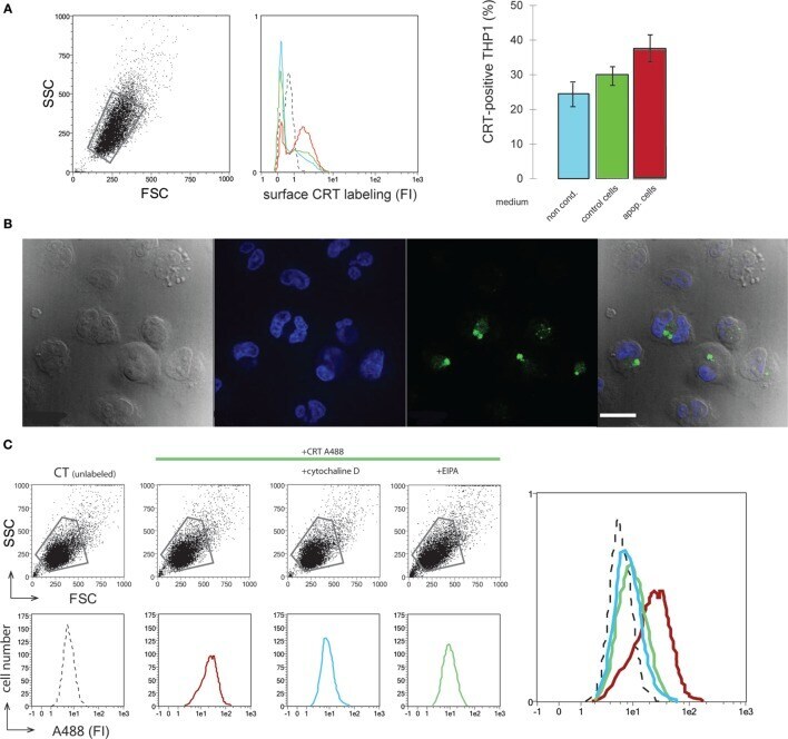
- Figure 3 Soluble calreticulin (CRT) binds to phagocyte and is endocytosed. (A) THP1 macrophages were incubated with normal medium (blue), medium conditioned by control (green), or apoptotic (red) JurkaT cells. Surface CRT was detected by immunobaleling and flow cytometry analysis. FSC/SSC dot plot, fluorescence histograms, and the quantification of the CRT-positive cells in the corresponding gated population were shown. Isotype control is shown in dotted line (middle panel). (B) THP1 macrophages incubated with CRT-A488 were visualized by microscopy under differential interference contrast, DAPI, and A488 filters, merge is shown (left to right panels). Bar: 22 um. (C) FSC/SSC dot plots and fluorescence histograms of the corresponding gated populations of THP1 macrophages incubated with CRT-A488 alone or in the presence of endocytic inhibitors as mentioned. An overlay of the data is shown. Unlabeled control cells (dotted line), CRT-A488 alone (red), CRT-A488 plus Cytochalasin D (light blue), and CRT-A488 plus 5-( N -Ethyl- N -isopropyl)amiloride (green).