 Explore
 Validate
 Learn
Learn Western blot
 ELISA
ELISAAntibody data
- Antibody Data
- Antigen structure
- References [0]
- Comments [0]
- Validations
- Western blot [2]
Submit
Validation data
Reference
Comment
Report error
- Product number
- NB100-2003-0.1mg - Provider product page

- Provider
- Novus Biologicals
- Product name
- Goat Polyclonal Aldolase A Antibody
- Antibody type
- Polyclonal
- Description
- Delipidation and Defibrination. This antibody will detect human Aldolase. Cross reactivity against Aldolase from other tissues and species may also occur. It has been reported that this antibody can detect human Aldolase on immunoblot showing a 41 kDa band in lysates from MCF7, NMB231 and HBL100 cell lines.
- Reactivity
- Human, Mouse, Rabbit
- Host
- Goat
- Conjugate
- Biotin
- Isotype
- IgG
- Vial size
- 0.1 mg
- Concentration
- LYOPH
- Storage
- Store at 4C short term. Aliquot and store at -20C long term. Avoid freeze-thaw cycles.
No comments: Submit comment
Supportive validation
- Submitted by
- Novus Biologicals (provider)
- Main image
- Experimental details
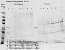
- Western Blot: Aldolase A Antibody [Biotin] [NB100-2003] - Immunoprecipitation was performed with 300 ul of anti aldolase antiserum and an equal volume of varied amounts (diluted from a stock solution of ~2.5 ug/ml) of purified aldolase in PBS. Precipitation of aldolase was confirmed by comparison of increasing amounts of antigen with the control protein by SDS PAGE and observation of a 40-45 kD MW band corresponding to Aldolase.

- Submitted by
- Novus Biologicals (provider)
- Main image
- Experimental details
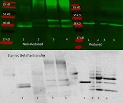
- Western Blot: Aldolase A Antibody [Biotin] [NB100-2003] - 300 ul aliquots of whole anti-aldolase antiserum were used to precipitate purified aldolase and precipitates with controls were compared by SDS-PAGE and Western blot. Samples shown are: 1. Purified aldolase 2. 300 ul antiserum, no antigen (neg control) 3. 300 ul antiserum with 100 ul aldolase (2.5 mg/ml) 4. 300 ul antiserum with 200 ul aldolase (2.5 mg/ml) For precipitation, 300 ul of antiserum and an equal volume of aldolase antigen in PBS was incubated 24 hrs at 4C, centrifuged 6 minutes at 13K RPM, washed once with PBS, centrifuged and dissolved in 60 ul 0.1 N NaOH. 90 ul of PBS added, the sample was divided in 2 portions, and an equal volume of reducing (+4% BME) or non-reducing 2X sample buffer was added. The reduced samples were boiled for five minutes, and all samples were run at 140 V for 45 minutes on a 4-20% tris/glycine gradient gel. Gel was stained, de-stained and imaged(see attached) using standard protocols.
