 Explore
 Validate
 Learn
Learn Western blot
 Immunoprecipitation
Antibody data
- Antibody Data
- Antigen structure
- References [0]
- Comments [0]
- Validations
- Immunoprecipitation [3]
Submit
Validation data
Reference
Comment
Report error
- Product number
- LS-C744807 - Provider product page

- Provider
- LSBio
- Product name
- ALDOA / Aldolase A Antibody (Liquid, Biotin) LS-C744807
- Antibody type
- Polyclonal
- Description
- Delipidation, salt fractionation and ion exchange chromatography followed by dialysis.
- Reactivity
- Human, Rabbit
- Host
- Goat
- Conjugate
- Biotin
- Isotype
- IgG
- Storage
- Store vial at -20°C or below prior to opening. Dilute 1:10 to minimize loss. Store the vial at -20°C or below after dilution. Avoid freeze-thaw cycles.
No comments: Submit comment
Supportive validation
- Submitted by
- LSBio (provider)
- Enhanced method
- Genetic validation
- Main image

- Experimental details
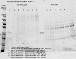
- Anti aldolase antibody– Immunoprecipitation- Immunoprecipitation was performed with 300 ul of anti aldolase antiserum and an equal volume of varied amounts (diluted from a stock solution of ~2.5 mg/ml) of purified aldolase in PBS. Antibody/Antigen mixture was incubated ~24 hrs at 4°C, centrifuged for 6 minutes at 13K RPM, washed once with PBS, centrifuged and dissolved in 60 ul 0.1 N NaOH. 90 ul of PBS was added, the sample was divided in 2 portions, and an equal volume of reducing (+4% BME) or non-reducing 2X sample buffer was added. The reduced samples were boiled for five minutes, and all samples were run at 140 V for ~45 minutes on a 4-20% tris/glycine gradient gel. Gel was stained, destained and imaged(see attached) using standard protocols. Precipitation of aldolase was confirmed by comparison of increasing amounts of antigen with the control protein by SDS PAGE and observation of a 40-45 kD MW band corresponding to Aldolase. Additional higher and lower molecular weight bands correspond to serum proteins.

- Submitted by
- LSBio (provider)
- Main image

- Experimental details
- Anti aldolase antibody – Immunoprecipitation and Western Blot. 300 µl aliquots of whole anti-aldolase antiserum (100-1141) were used to precipitate varying amounts of purified aldolase and precipitates with controls were compared by SDS-PAGE and Western blot. Samples shown in the image are: 1. Purified aldolase 2. 300 µl antiserum with no antigen (negative control) 3. 300 µl antiserum with ~100 µl aldolase (2.5 mg/ml) 4. 300 µl antiserum with ~200 µl aldolase (2.5 mg/ml) For the precipitation, 300 ul of antiserum and an equal volume of aldolase antigen in PBS was incubated ~24 hrs at 4°C, centrifuged for 6 minutes at 13K RPM, washed once with PBS, centrifuged and dissolved in 60 ul 0.1 N NaOH. 90 ul of PBS was added, the sample was divided in 2 portions, and an equal volume of reducing (+4% BME) or non-reducing 2X sample buffer was added. The reduced samples were boiled for five minutes, and all samples were run at 140 V for ~45 minutes on a 4-20% tris/glycine gradient gel. Gel was stained, destained and imaged(see attached) using standard protocols. Precipitation of aldolase was confirmed by comparison of increasing amounts of antigen with the control protein by SDS PAGE and observation of a 40-45 kD MW band corresponding to Aldolase. Additional higher and lower molecular weight bands correspond to serum proteins.

- Submitted by
- LSBio (provider)
- Main image
- Experimental details
- Anti aldolase antibody– Immunoprecipitation- Immunoprecipitation was performed with 300 ul of anti aldolase antiserum and an equal volume of varied amounts (diluted from a stock solution of ~2.5 mg/ml) of purified aldolase in PBS. Antibody/Antigen mixture was incubated ~24 hrs at 4°C, centrifuged for 6 minutes at 13K RPM, washed once with PBS, centrifuged and dissolved in 60 ul 0.1 N NaOH. 90 ul of PBS was added, the sample was divided in 2 portions, and an equal volume of reducing (+4% BME) or non-reducing 2X sample buffer was added. The reduced samples were boiled for five minutes, and all samples were run at 140 V for ~45 minutes on a 4-20% tris/glycine gradient gel. Gel was stained, destained and imaged(see attached) using standard protocols. Precipitation of aldolase was confirmed by comparison of increasing amounts of antigen with the control protein by SDS PAGE and observation of a 40-45 kD MW band corresponding to Aldolase. Additional higher and lower molecular weight bands correspond to serum proteins.
