Explore
 Validate
 Learn
Learn Western blot
 ELISA
ELISA Immunoprecipitation
Antibody data
- Antibody Data
- Antigen structure
- References [0]
- Comments [0]
- Validations
- Western blot [1]
- Other assay [2]
Submit
Validation data
Reference
Comment
Report error
- Product number
- 100-1141 - Provider product page

- Provider
- Invitrogen Antibodies
- Product name
- Aldolase Polyclonal Antibody
- Antibody type
- Polyclonal
- Antigen
- Other
- Reactivity
- Human, Rabbit
- Host
- Goat
- Isotype
- IgG
- Vial size
- 2 mL
- Concentration
- 90 mg/mL
- Storage
- Store at 4°C short term. For long term storage, store at -20°C, avoiding freeze/thaw cycles.
No comments: Submit comment
Supportive validation
- Submitted by
- Invitrogen Antibodies (provider)
- Main image
- Experimental details
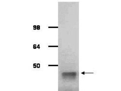
- IgG purified antibody to rabbit muscle aldolase (100-1141, 200-1141 and 200-1341) was used at a 1:1000 dilution to detect human aldolase by Western blot. A whole cell lysate prepared from human derived A293 cells was loaded on a 4-12% tris glycine gradient gel for SDS-PAGE. The gel was transferred to nitro-cellulose using standard techniques. Antibody reaction with the membrane occurred overnight at 4° C in TTBS supplemented with 2% non-fat dry milk. Color was allowed to develop using SuperSignal West Pico Chemiluminescent Substrate (PIERCE). Other detection methods will yield similar results. This antibody clearly detects a band at ~41 kDa consistent with human aldolase.

Supportive validation
- Submitted by
- Invitrogen Antibodies (provider)
- Main image
- Experimental details
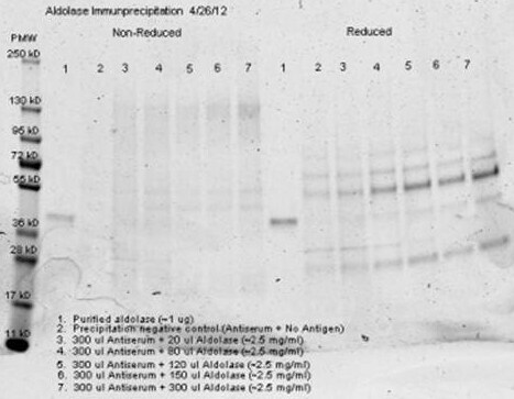
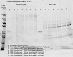
- Anti aldolase antibody - immunoprecipitation and western blot. 300 µl aliquots of whole anti-aldolase antiserum (100-1141) were used to precipitate varying amounts of purified aldolase and precipitates with controls were compared by SDS-PAGE and Western blot. Samples shown in the image are: 1. Purified aldolase 2. 300 µl antiserum with no antigen (negative control) 3. 300 µl antiserum with ~100 µl aldolase (2.5 mg/ml) 4. 300 µl antiserum with ~200 µl aldolase (2.5 mg/ml) For the precipitation, 300 ul of antiserum and an equal volume of aldolase antigen in PBS was incubated ~24 hrs at 4°C, centrifuged for 6 minutes at 13K RPM, washed once with PBS, centrifuged and dissolved in 60 ul 0.1 N NaOH. 90 ul of PBS was added, the sample was divided in 2 portions, and an equal volume of reducing (+4% BME) or non-reducing 2X sample buffer was added. The reduced samples were boiled for five minutes, and all samples were run at 140 V for ~45 minutes on a 4-20% tris/glycine gradient gel. Gel was stained, destained and imaged(see attached) using standard protocols. Precipitation of aldolase was confirmed by comparison of increasing amounts of antigen with the control protein by SDS PAGE and observation of a 40-45 kDa MW band corresponding to Aldolase. Additional higher and lower molecular weight bands correspond to serum proteins.
- Submitted by
- Invitrogen Antibodies (provider)
- Main image
- Experimental details
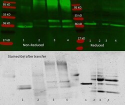
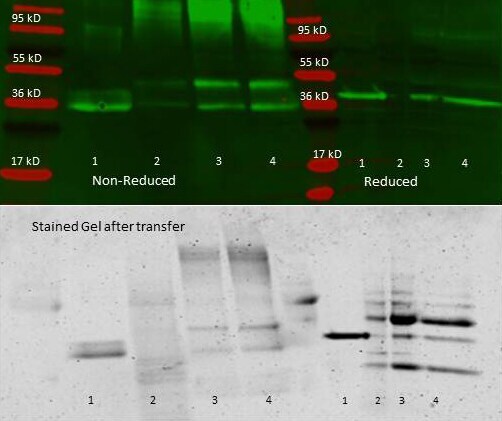
- Immunoprecipitation of rabbit anti Aldolase antiserum - Immunoprecipitation performed with 300 ul of antiserum and an equal volume of varied amounts of purified aldolase diluted from a stock solution of ~2.5 mg/ml aldolase in PBS. Antibody/Antigen mixture was incubated ~24 hrs at 4°C, centrifuged for 6 minutes at 13K RPM, washed once with PBS, centrifuged and dissolved in 60 ul 0.1 N NaOH. 90 ul of PBS was added, the sample was divided in 2 portions, and an equal volume of reducing (+4% BME) or non-reducing 2X sample buffer was added. The reduced samples were boiled for five minutes, and all samples were run at 140 V for ~45 minutes on a 4-20% tris/glycine gradient gel. Gel was stained, destained and imaged (see attached) using standard protocols. Precipitation of aldolase was confirmed by comparison of increasing amounts of antigen with the control protein by SDS PAGE and observation of a 40-45 kDa MW band corresponding to Aldolase. Additional higher and lower molecular weight bands correspond to serum proteins.