Explore
 Validate
 Learn
Learn Western blot
 Immunocytochemistry
Antibody data
- Antibody Data
- Antigen structure
- References [9]
- Comments [0]
- Validations
- Immunocytochemistry [1]
Submit
Validation data
Reference
Comment
Report error
- Product number
- HPA004177 - Provider product page

- Provider
- Atlas Antibodies
- Proper citation
- Atlas Antibodies Cat#HPA004177, RRID:AB_1078128
- Product name
- Anti-ALDOA
- Antibody type
- Polyclonal
- Description
- Polyclonal Antibody against Human ALDOA, Gene description: aldolase A, fructose-bisphosphate, Validated applications: ICC, IHC, WB, Uniprot ID: P04075, Storage: Store at +4°C for short term storage. Long time storage is recommended at -20°C.
- Reactivity
- Human
- Host
- Rabbit
- Conjugate
- Unconjugated
- Isotype
- IgG
- Vial size
- 100 µl
- Concentration
- 0.05 mg/ml
- Storage
- Store at +4°C for short term storage. Long time storage is recommended at -20°C.
- Handling
- The antibody solution should be gently mixed before use.
Submitted references The fructose-bisphosphate, Aldolase A (ALDOA), facilitates DNA-PKcs and ATM kinase activity to regulate DNA double-strand break repair
A Proximity biotinylation assay with a host protein bait reveals multiple factors modulating enterovirus replication
The ALDOA Metabolism Pathway as a Potential Target for Regulation of Prostate Cancer Proliferation
Expression of Genes in the 16p11.2 Locus during Development of the Human Fetal Cerebral Cortex
Glycolytic biomarkers predict transformation in patients with follicular lymphoma.
High expression of Aldolase A predicts poor survival in patients with clear-cell renal cell carcinoma
Differentially expressed proteins in malignant and benign adrenocortical tumors.
Comparative Proteomics Analysis of Gastric Cancer Stem Cells
Fructose-Bisphosphate Aldolase A Is a Potential Metastasis-Associated Marker of Lung Squamous Cell Carcinoma and Promotes Lung Cell Tumorigenesis and Migration
Sobanski T, Suraweera A, Burgess J, Richard I, Cheong C, Dave K, Rose M, Adams M, O’Byrne K, Richard D, Bolderson E
Scientific Reports 2023;13(1)
Scientific Reports 2023;13(1)
A Proximity biotinylation assay with a host protein bait reveals multiple factors modulating enterovirus replication
Semler B, Moghimi S, Viktorova E, Gabaglio S, Zimina A, Budnik B, Wynn B, Sztul E, Belov G
PLOS Pathogens 2022;18(10):e1010906
PLOS Pathogens 2022;18(10):e1010906
The ALDOA Metabolism Pathway as a Potential Target for Regulation of Prostate Cancer Proliferation
Kuang Q, Liang Y, Zhuo Y, Cai Z, Jiang F, Xie J, Zheng Y, Zhong W
OncoTargets and Therapy 2021;Volume 14
OncoTargets and Therapy 2021;Volume 14
Expression of Genes in the 16p11.2 Locus during Development of the Human Fetal Cerebral Cortex
Morson S, Yang Y, Price D, Pratt T
Cerebral Cortex 2021;31(9):4038-4052
Cerebral Cortex 2021;31(9):4038-4052
Glycolytic biomarkers predict transformation in patients with follicular lymphoma.
Monrad I, Madsen C, Lauridsen KL, Honoré B, Plesner TL, Hamilton-Dutoit S, d'Amore F, Ludvigsen M
PloS one 2020;15(5):e0233449
PloS one 2020;15(5):e0233449
High expression of Aldolase A predicts poor survival in patients with clear-cell renal cell carcinoma
Na N, Li H, Xu C, Bin M, Hong L, Huang Z, Qiu J
Therapeutics and Clinical Risk Management 2017;Volume 13
Therapeutics and Clinical Risk Management 2017;Volume 13
Differentially expressed proteins in malignant and benign adrenocortical tumors.
Kjellin H, Johansson H, Höög A, Lehtiö J, Jakobsson PJ, Kjellman M
PloS one 2014;9(2):e87951
PloS one 2014;9(2):e87951
Comparative Proteomics Analysis of Gastric Cancer Stem Cells
Hjelmeland A, Morisaki T, Yashiro M, Kakehashi A, Inagaki A, Kinoshita H, Fukuoka T, Kasashima H, Masuda G, Sakurai K, Kubo N, Muguruma K, Ohira M, Wanibuchi H, Hirakawa K
PLoS ONE 2014;9(11):e110736
PLoS ONE 2014;9(11):e110736
Fructose-Bisphosphate Aldolase A Is a Potential Metastasis-Associated Marker of Lung Squamous Cell Carcinoma and Promotes Lung Cell Tumorigenesis and Migration
Singh S, Du S, Guan Z, Hao L, Song Y, Wang L, Gong L, Liu L, Qi X, Hou Z, Shao S
PLoS ONE 2014;9(1):e85804
PLoS ONE 2014;9(1):e85804
No comments: Submit comment
Supportive validation
- Submitted by
- Atlas Antibodies (provider)
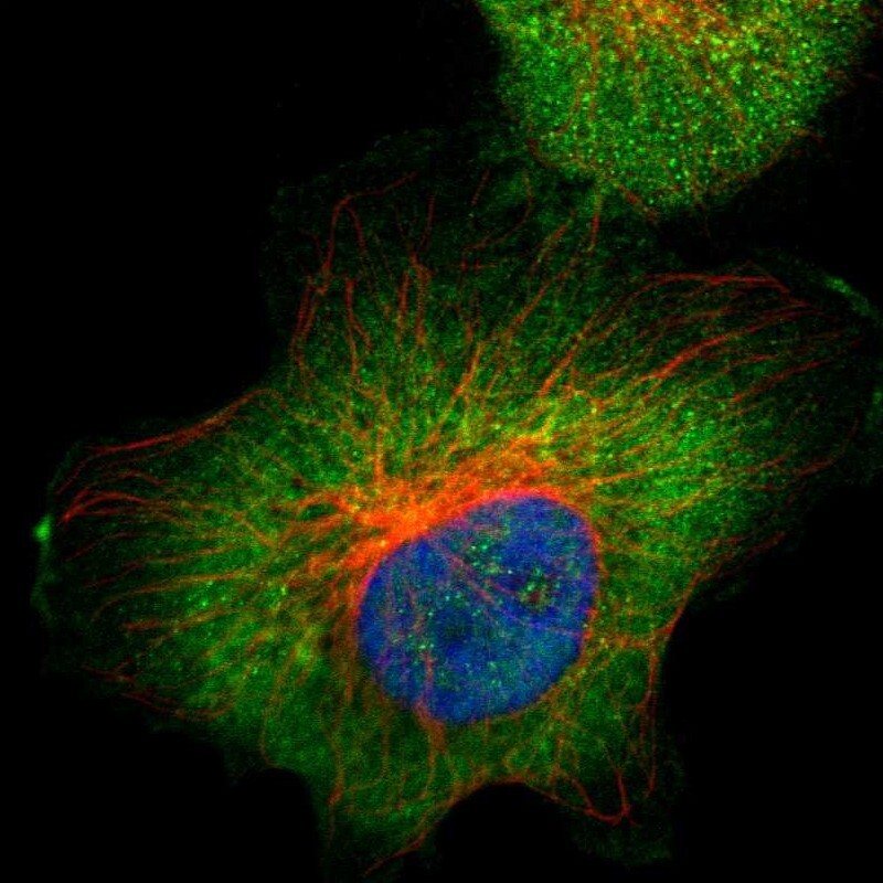
- Main image
- Experimental details
- Immunofluorescent staining of human cell line U-251 MG shows localization to cytosol.
- Sample type
- Human