Explore
 Validate
 Learn
Learn Immunocytochemistry
Antibody data
- Antibody Data
- Antigen structure
- References [0]
- Comments [0]
- Validations
- Immunocytochemistry [6]
- Immunohistochemistry [6]
Submit
Validation data
Reference
Comment
Report error
- Product number
- PA5-55721 - Provider product page

- Provider
- Invitrogen Antibodies
- Product name
- NUP153 Polyclonal Antibody
- Antibody type
- Polyclonal
- Antigen
- Recombinant protein fragment
- Description
- Immunogen sequence: SSRASDKDIT VSKNTSLPPL WSPEAERSHS LSQHTATSSK KPAFNLSAFG TLSPSLGNSS ILKTSQLGDS PFYPGKTTYG GAAAAVRQSK LRNTP Highest antigen sequence identity to the following orthologs: Mouse - 93%, Rat - 93%.
- Reactivity
- Human
- Host
- Rabbit
- Isotype
- IgG
- Vial size
- 100 μL
- Concentration
- 0.1 mg/mL
- Storage
- Store at 4°C short term. For long term storage, store at -20°C, avoiding freeze/thaw cycles.
No comments: Submit comment
Supportive validation
- Submitted by
- Invitrogen Antibodies (provider)
- Main image
- Experimental details
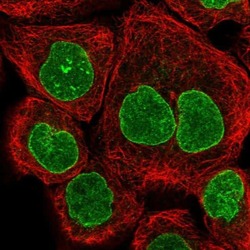
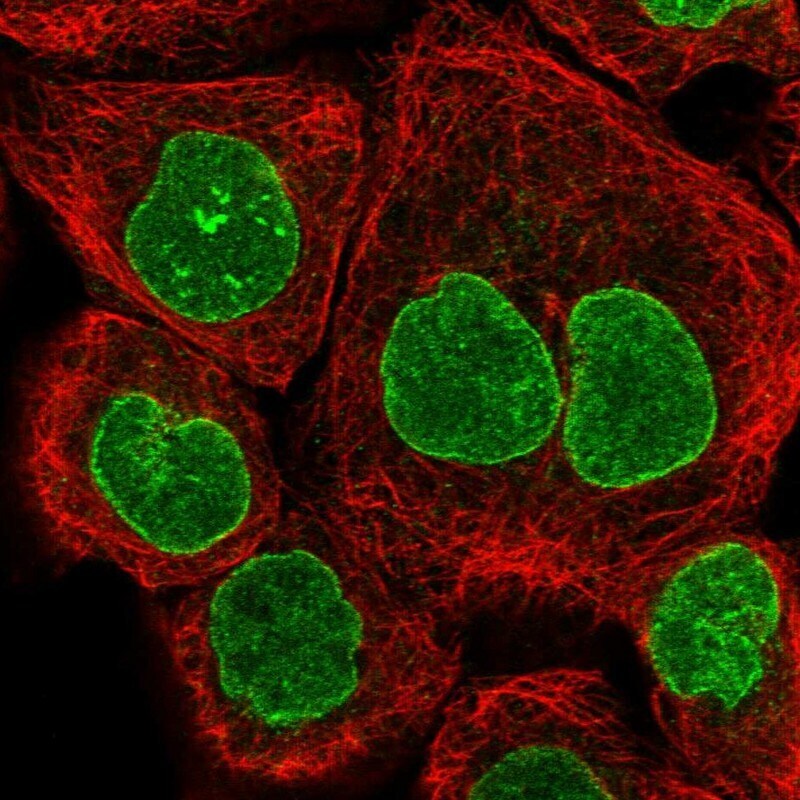
- Immunofluorescent staining of NUP153 in human cell line A-431 shows positivity in nuclear membrane. Samples were probed using a NUP153 Polyclonal Antibody (Product # PA5-55721).

- Submitted by
- Invitrogen Antibodies (provider)
- Main image
- Experimental details
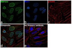
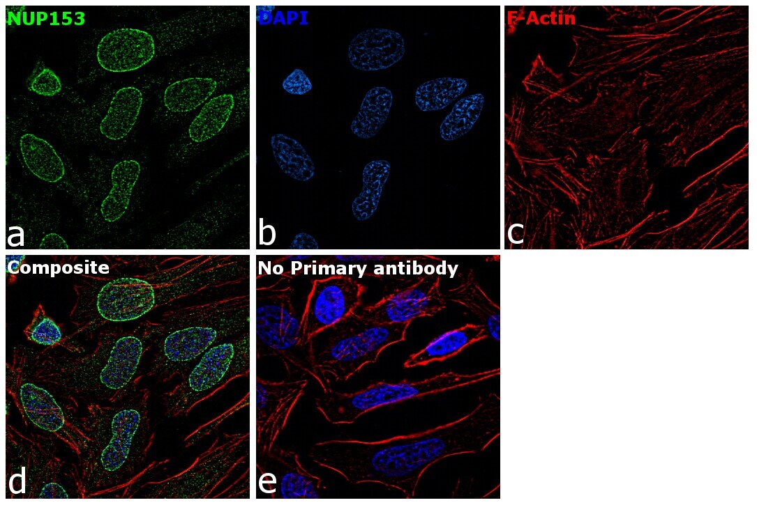
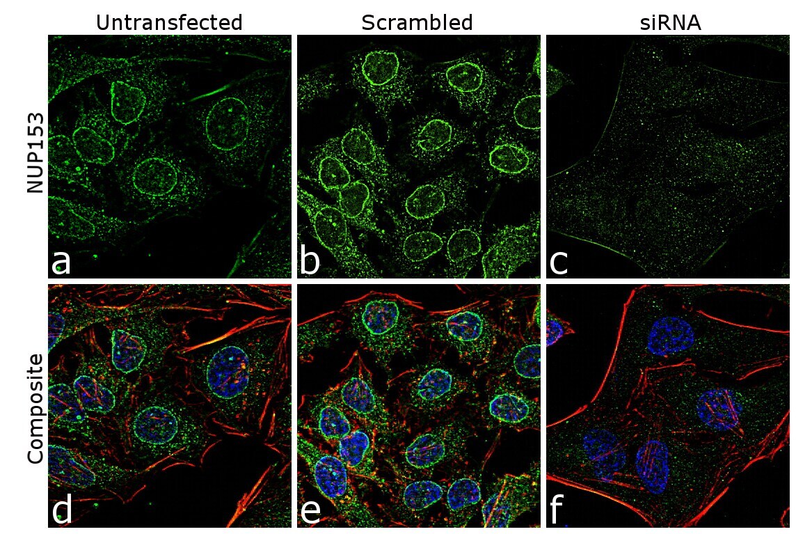
- Immunofluorescence analysis of NUP153 was performed using 70% confluent log phase HeLa cells. The cells were fixed with 4% paraformaldehyde for 10 minutes, permeabilized with 0.1% Triton™ X-100 for 15 minutes, and blocked with 2% BSA for 1 hour at room temperature. The cells were labeled with NUP153 Rabbit Polyclonal Antibody (Product # PA5-55721) at 5 µg/mL in 0.1% BSA, incubated at 4 degree Celsius overnight and then labeled with Goat anti-Rabbit IgG (H+L) Superclonal™ Secondary Antibody, Alexa Fluor® 488 conjugate (Product # A27034) at a dilution of 1:2000 for 45 minutes at room temperature (Panel a: green). Nuclei (Panel b: blue) were stained with ProLong™ Diamond Antifade Mountant with DAPI (Product # P36962). F-actin (Panel c: red) was stained with Rhodamine Phalloidin (Product # R415). Panel d represents the merged image showing Nuclear and Nuclear membrane localization. Panel e represents control cells with no primary antibody to assess background. The images were captured at 60X magnification.
- Submitted by
- Invitrogen Antibodies (provider)
- Main image
- Experimental details
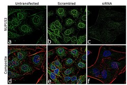
- Knockdown of NUP153 was achieved by transfecting HeLa cells with NUP153 specific siRNA (Silencer® select Product # s19374). Immunofluorescence analysis was performed on HeLa cells (untransfected, panel a,d), transfected with non-specific scrambled siRNA (panels b,e) and transfected with NUP153 specific siRNA (panel c,f). Cells were fixed, permeabilized, and labelled with NUP153 Rabbit Polyclonal Antibody (Product # PA5-55721, 5 µg/mL), followed by Goat anti-Rabbit IgG (H+L) Superclonal™ Secondary Antibody, Alexa Fluor® 488 conjugate (Product # A27034, 1:2000). Nuclei (blue) were stained using ProLong™ Diamond Antifade Mountant with DAPI (Product # P36962), and Rhodamine Phalloidin (Product # R415, 1:300) was used for cytoskeletal F-actin (red) staining. Reduction of specific signal was observed upon siRNA mediated knockdown (panel c,f) confirming specificity of the antibody to NUP153 (green). The images were captured at 60X magnification.
- Submitted by
- Invitrogen Antibodies (provider)
- Main image

- Experimental details
- Knockdown of NUP153 was achieved by transfecting HeLa cells with NUP153 specific siRNA (Silencer® select Product # s19374). Immunofluorescence analysis was performed on HeLa cells (untransfected, panel a,d), transfected with non-specific scrambled siRNA (panels b,e) and transfected with NUP153 specific siRNA (panel c,f). Cells were fixed, permeabilized, and labelled with NUP153 Rabbit Polyclonal Antibody (Product # PA5-55721, 5 µg/mL), followed by Goat anti-Rabbit IgG (Heavy Chain) Superclonal™ Secondary Antibody, Alexa Fluor® 488 conjugate (Product # A27034, 1:2000). Nuclei (blue) were stained using ProLong™ Diamond Antifade Mountant with DAPI (Product # P36962), and Rhodamine Phalloidin (Product # R415, 1:300) was used for cytoskeletal F-actin (red) staining. Reduction of specific signal was observed upon siRNA mediated knockdown (panel c,f) confirming specificity of the antibody to NUP153 (green). The images were captured at 60X magnification.

- Submitted by
- Invitrogen Antibodies (provider)
- Main image

- Experimental details
- Immunofluorescence analysis of NUP153 was performed using 70% confluent log phase HeLa cells. The cells were fixed with 4% paraformaldehyde for 10 minutes, permeabilized with 0.1% Triton™ X-100 for 15 minutes, and blocked with 2% BSA for 1 hour at room temperature. The cells were labeled with NUP153 Rabbit Polyclonal Antibody (Product # PA5-55721) at 5 µg/mL in 0.1% BSA, incubated at 4 degree Celsius overnight and then labeled with Goat anti-Rabbit IgG (Heavy Chain) Superclonal™ Secondary Antibody, Alexa Fluor® 488 conjugate (Product # A27034) at a dilution of 1:2000 for 45 minutes at room temperature (Panel a: green). Nuclei (Panel b: blue) were stained with ProLong™ Diamond Antifade Mountant with DAPI (Product # P36962). F-actin (Panel c: red) was stained with Rhodamine Phalloidin (Product # R415). Panel d represents the merged image showing Nuclear and Nuclear membrane localization. Panel e represents control cells with no primary antibody to assess background. The images were captured at 60X magnification.

- Submitted by
- Invitrogen Antibodies (provider)
- Main image

- Experimental details
- Immunofluorecent analysis of NUP153 in human cell line A-431 using NUP153 Polyclonal Antibody (Product # PA5-55721). Staining shows localization to nuclear membrane.
Supportive validation
- Submitted by
- Invitrogen Antibodies (provider)
- Main image
- Experimental details
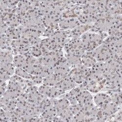
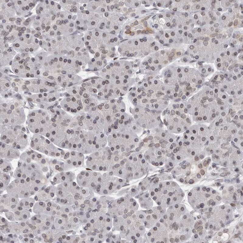
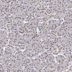
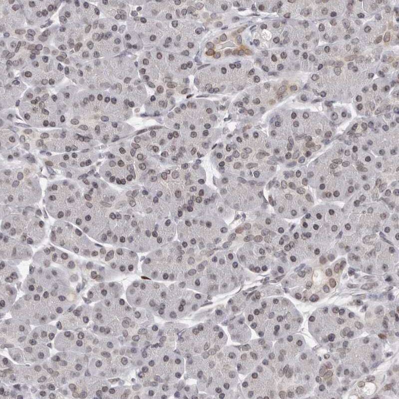
- Immunohistochemical staining of NUP153 in human pancreas using NUP153 Polyclonal Antibody (Product # PA5-55721) shows very weak positivity in nuclear membrane in exocrine glandular cells.
- Submitted by
- Invitrogen Antibodies (provider)
- Main image
- Experimental details
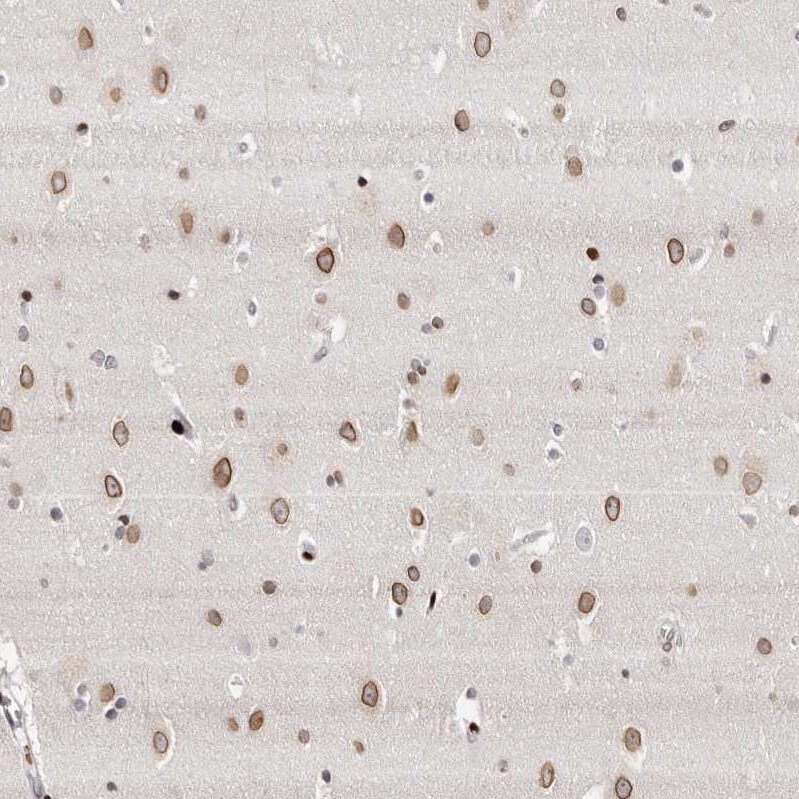
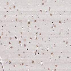
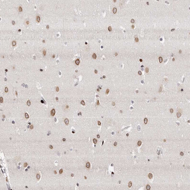
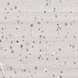
- Immunohistochemical staining of NUP153 in human cerebral cortex using NUP153 Polyclonal Antibody (Product # PA5-55721) shows moderate to strong positivity in nuclear membrane in neurons.
- Submitted by
- Invitrogen Antibodies (provider)
- Main image
- Experimental details
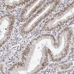
- Immunohistochemical staining of NUP153 in human endometrium using NUP153 Polyclonal Antibody (Product # PA5-55721) shows weak to moderate positivity in nuclear membrane in glandular cells.

- Submitted by
- Invitrogen Antibodies (provider)
- Main image
- Experimental details
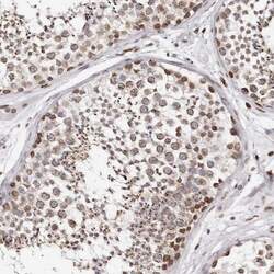
- Immunohistochemical staining of NUP153 in human testis using NUP153 Polyclonal Antibody (Product # PA5-55721) shows moderate positivity in nuclear membrane in cells in seminiferous ducts.

- Submitted by
- Invitrogen Antibodies (provider)
- Main image
- Experimental details
- Immunohistochemical staining of NUP153 in human pancreas using NUP153 Polyclonal Antibody (Product # PA5-55721) shows very weak positivity in nuclear membrane in exocrine glandular cells.
- Submitted by
- Invitrogen Antibodies (provider)
- Main image
- Experimental details
- Immunohistochemical staining of NUP153 in human cerebral cortex using NUP153 Polyclonal Antibody (Product # PA5-55721) shows moderate to strong positivity in nuclear membrane in neurons.