Explore
 Validate
 Learn
Learn Western blot
Antibody data
- Antibody Data
- Antigen structure
- References [0]
- Comments [0]
- Validations
- Western blot [2]
- Immunocytochemistry [2]
Submit
Validation data
Reference
Comment
Report error
- Product number
- PA5-66752 - Provider product page

- Provider
- Invitrogen Antibodies
- Product name
- beta-1 Adaptin Polyclonal Antibody
- Antibody type
- Polyclonal
- Antigen
- Recombinant full-length protein
- Description
- Immunogen sequence: PPSAFVEGGRG VVHKSLPPRT ASSESAESPE TAPTGAPP
- Concentration
- 0.7 mg/mL
No comments: Submit comment
Supportive validation
- Submitted by
- Invitrogen Antibodies (provider)
- Main image
- Experimental details
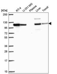
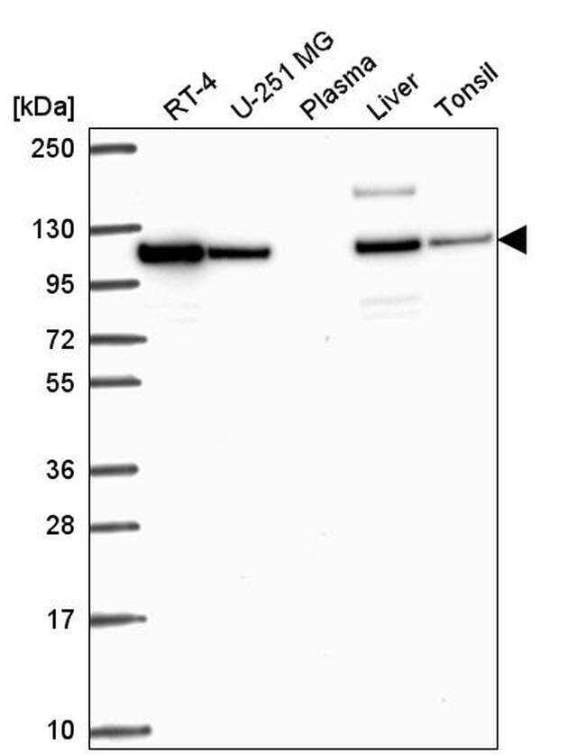
- Western blot analysis of beta-1 Adaptin in human cell line RT-4, human cell line U-251 MG, human plasma, human liver tissue and human tonsil tissue. Samples were probed using a beta-1 Adaptin Polyclonal Antibody (Product # PA5-66752).
- Submitted by
- Invitrogen Antibodies (provider)
- Main image
- Experimental details
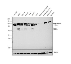
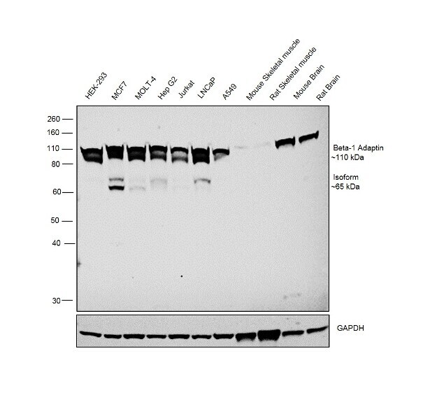
- Western blot was performed using anti-beta-1 Adaptin Polyclonal Antibody (Product # PA5-66752) and a 110kDa band corresponding to beta-1 Adaptin was observed across all the cell lines and tissue lysates tested except Mouse Skeletal Muscle and Rat Skeletal Muscle which are reported negative for beta-1 Adaptin expression. An additional band which corresponds to an isoform of beta-1 Adaptin was detected at 65KDa in MCF7, MOLT-4, HepG2, Jurkat and LNCaP. Whole cell extracts (30 µg lysate) of HEK-293 (Lane 1), MCF7 (Lane 2), MOLT-4 (Lane 3), Hep G2 (Lane 4), Jurkat (Lane 5), LNCaP (Lane 6), A549 (Lane 7), tissue extracts of Mouse Skeletal Muscle (Lane 8), Rat Skeletal Muscle (Lane 9), Mouse Brain (Lane 10) and Rat Brain (Lane 11) were electrophoresed using NuPAGE® 10 % Bis-Tris gel (Product # NP0302BOX). Resolved proteins were then transferred onto a nitrocellulose membrane (Product # IB23001) by iBlot® 2 Dry Blotting System (Product # IB21001). The blots were probed with the primary antibody (1:200 dilution) and detected by chemiluminescence Goat Anti-Rabbit IgG Secondary Antibody, HRP conjugate (Product # A27036, 1:4000 dilution) using the iBright FL 1000 (Product # A32752). Chemiluminescent detection was performed using Novex® ECL Chemiluminescent Substrate Reagent Kit (Product # WP20005).
Supportive validation
- Submitted by
- Invitrogen Antibodies (provider)
- Main image
- Experimental details
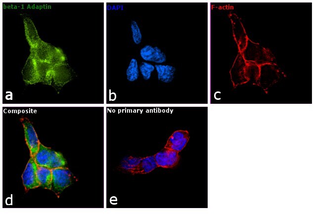
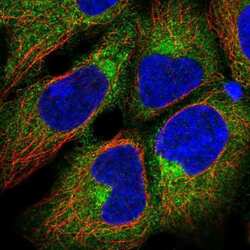
- Immunofluorescent staining of beta-1 Adaptin in human cell line HEK 293 shows localization to cytosol and the Golgi apparatus. Samples were probed using a beta-1 Adaptin Polyclonal Antibody (Product # PA5-66752).

- Submitted by
- Invitrogen Antibodies (provider)
- Main image

- Experimental details
- Immunofluorescence analysis of beta-1 Adaptin was performed using 70% confluent log phase HEK-293 cells. The cells were fixed with 4% paraformaldehyde for 10 minutes, permeabilized with 0.1% Triton™ X-100 for 15 minutes, and blocked with 1% BSA for 1 hour at room temperature. The cells were labeled with beta-1 Adaptin Polyclonal Antibody (Product # PA5-66752) at 1:100 dilution in 0.1% BSA, incubated at 4 degree Celsius overnight and then labeled with Goat anti-Rabbit IgG (H+L) Superclonal™ Secondary Antibody, Alexa Fluor® 488 conjugate (Product # A27034) at a dilution of 1:2000 for 45 minutes at room temperature (Panel a: green). Nuclei (Panel b: blue) were stained with SlowFade® Gold Antifade Mountant with DAPI (Product # S36938). F-actin (Panel c: red) was stained with Rhodamine Phalloidin (Product # R415, 1:300). Panel d represents the merged image showing cytoplasmic, golgi and vesicular localization. Panel e represents control cells with no primary antibody to assess background. The images were captured at 60X magnification. .