Explore
 Validate
 Learn
Learn Western blot
Antibody data
- Antibody Data
- Antigen structure
- References [0]
- Comments [0]
- Validations
- Western blot [1]
- Immunocytochemistry [1]
- Immunohistochemistry [1]
Submit
Validation data
Reference
Comment
Report error
- Product number
- TA328849 - Provider product page

- Provider
- OriGene
- Product name
- Rabbit Polyclonal Anti-Galanin Receptor Type 1
- Antibody type
- Polyclonal
- Description
- Rabbit Polyclonal Anti-Galanin Receptor Type 1
- Host
- Rabbit
- Conjugate
- Unconjugated
- Epitope
- Galr1
- Antibody clone number
- NULL
- Vial size
- 200 µl
- Concentration
- NULL
No comments: Submit comment
Supportive validation
- Submitted by
- OriGene (provider)
- Main image
- Experimental details
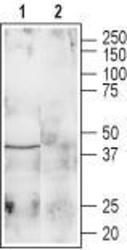
- Western blot analysis of rat brain lysate: 1. Anti-Galanin Receptor type 1 antibody, (1:200). 2. Anti-Galanin Receptor type 1 antibody, preincubated with the control peptide antigen.
- Validation comment
- WB

Supportive validation
- Submitted by
- OriGene (provider)
- Main image
- Experimental details
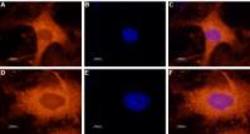
- Expression of GALR1 in rat DRG. Immunocytochemical staining of GALR1 in a primary culture of rat dorsal root ganglion (DRG) neurons. A, D. Paraformaldehyde-fixed and permeabilized DRG primary culture stained with Anti-Galanin Receptor Type 1 antibody, (1:100) followed by goat-anti-rabbit-AlexaFluor-555 secondary antibody (red). B, E. Nuclear fluorescence staining of cells using Hoechst 33342 (blue). C. Merged images of panels A and B. F. Merged images of panels D and E.
- Validation comment
- IF

Supportive validation
- Submitted by
- OriGene (provider)
- Main image

- Experimental details
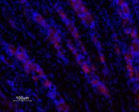
- Expression of GALR1 in DRG. Immunohistochemical staining of rat dorsal root ganglia (DRG) frozen section using Anti-Galanin Receptor Type 1 antibody, (1:200), (red). Galanin Receptor is expressed in DRG neuron cells. Hoechst 33342 is used as the counterstain (blue).
- Validation comment
- IHC